The Structure of Skeletal Muscle (AQA A Level Biology): Revision Note
Exam code: 7402

Skeletal muscle structure
Skeletal muscle, also known as striated, or striped, muscle, is the muscle that contracts to move the skeleton
Other types of muscle include cardiac muscle in the heart, and smooth muscle in the walls of the internal organs
Skeletal muscle is made up of bundles of muscle cells
Muscle cells are usually known as muscle fibres
In a muscle fibre the:
cell surface membrane = sarcolemma
cytoplasm = sarcoplasm
endoplasmic reticulum = sarcoplasmic reticulum
Muscle fibres have the following features:
each muscle fibre is an elongated cell that contains many nuclei
the sarcoplasm contains an organised arrangement of contractile proteins that form myofibrils
the sarcolemma has many deep tube-like projections that fold in from its outer surface; these are known as T-tubules
many mitochondria in the sarcoplasm generate ATP for muscle contraction
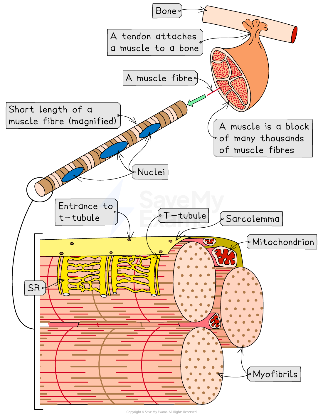
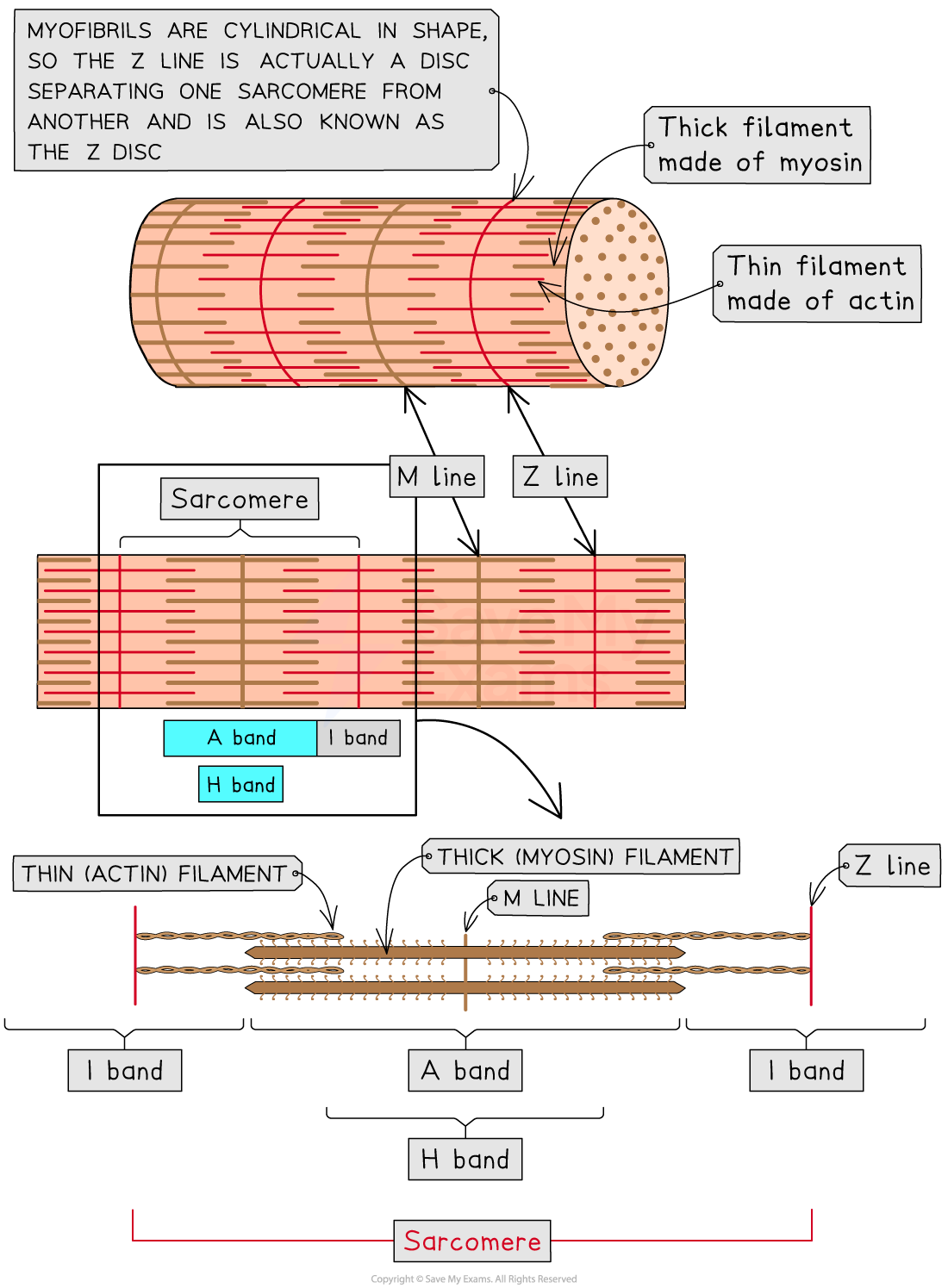
Myofibrils
Myofibrils are long, rod-like structures made of protein filaments
The protein filaments found in myofibrils are:
myosin, also known as thick filaments
actin, also known as thin filaments
Myofibrils can be divided up into sections known as sarcomeres, which shorten during muscle contraction as myosin and actin filaments slide past each other
This is the sliding filament model of muscle contraction
Each sarcomere contains the following features:
Feature | Description |
|---|---|
Z-line | The boundary between sarcomeres Actin filaments attach here During muscle contraction the z-lines get closer together |
M-line | The central point of each sarcomere Myosin filaments attach here |
A band | The region of the sarcomere across which the myosin filaments extend Myosin filaments do not change in length, so the A band remains the same size whether the muscle is contracted or relaxed |
H band | The region of the sarcomere containing only myosin filaments The H band shrinks during muscle contraction as the overlap between actin and myosin filaments increases |
I band | The region of the sarcomere containing only actin filaments The I band shrinks during muscle contraction as the overlap between actin and myosin filaments increases |
Examiner Tips and Tricks
There is a lot of complicated language associated with muscle structure, so be sure that you can define the important key words; common pitfalls include confusion between:
myofibril (muscles) with microfibrils (cellulose)
muscle fibres and protein filaments
myofibrils and sarcomeres
You should also make sure that you know how sarcomeres change during muscle contraction:
The A band remains the same length
The H band and I band become shorter
The Z lines get closer together

Unlock more, it's free!
Join the 100,000+ Students that ❤️ Save My Exams
the (exam) results speak for themselves:

Was this revision note helpful?
Build on this topic