11 mark
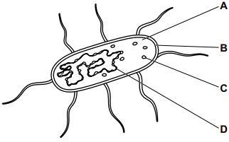
Which label represents a plasmid in this diagram of a bacterium?
Was this exam question helpful?
Exam code: 0653
Which label represents a plasmid in this diagram of a bacterium?
Was this exam question helpful?
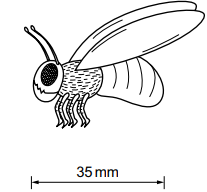
The actual length of a phorid fly is 5.5 mm.
What is the magnification of this diagram of a phorid fly?
0.16
0.7
6.4
192.5
Was this exam question helpful?
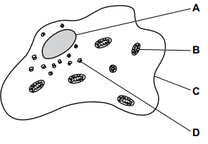
Which label shows the site of aerobic respiration in this diagram of an animal cell?
Was this exam question helpful?
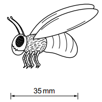
The actual length of a phorid fly is 5500 μm long.
What is the magnification of this diagram of a phorid fly?
0.006
0.16
6.4
157
Was this exam question helpful?
What is a type of tissue in a living organism?
blood
heart
plasma
plasmid
Was this exam question helpful?
Fig. 1.1 shows an animal cell and a plant cell.
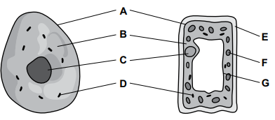
Fig 1.1
Table 1.1 shows the names and functions of some of the structures of the cells labelled in Fig. 1.1.
letter in Fig. 1.1 | name of structure | function |
A | ...................... | controls the movement of substances into and out of cells |
D | mitochondria | ...................... |
.......... | vacuole | ...................... |
Fig. 1.2 shows some structures organised in order of increasing size, starting with the smallest size.
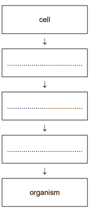
Fig. 1.2
Use the words from the list to complete Fig. 1.2.
organ organ system tissue
Was this exam question helpful?