Recognising Structures (Cambridge (CIE) A Level Biology): Revision Note

Exam code: 9700

Emma Archbold

Written by: Emma Archbold

Reviewed by: Alistair Marjot

Updated on

Recognising structures in the gas exchange system

Trachea

  • A tracheal cross-section shows the large lumen, which air has to travel through

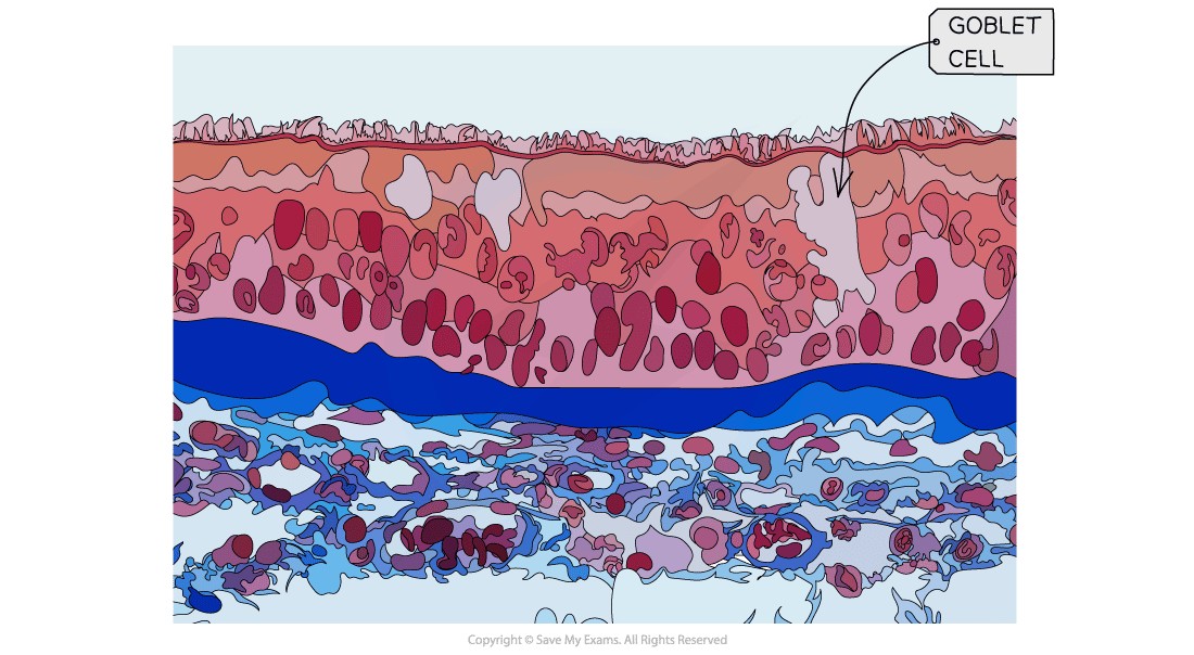
  • The innermost cells of the trachea are the ciliated epithelia with projections called cilia

Trachea cross-section as shown under a light microscope
  • The cells of the ciliated epithelium are shown in the light micrograph below

    • The cells are tightly packed and interspersed with goblet cells, which are shown in light grey

A light micrograph image showing tissue of the trachea
A photomicrograph image showing tissue of the trachea
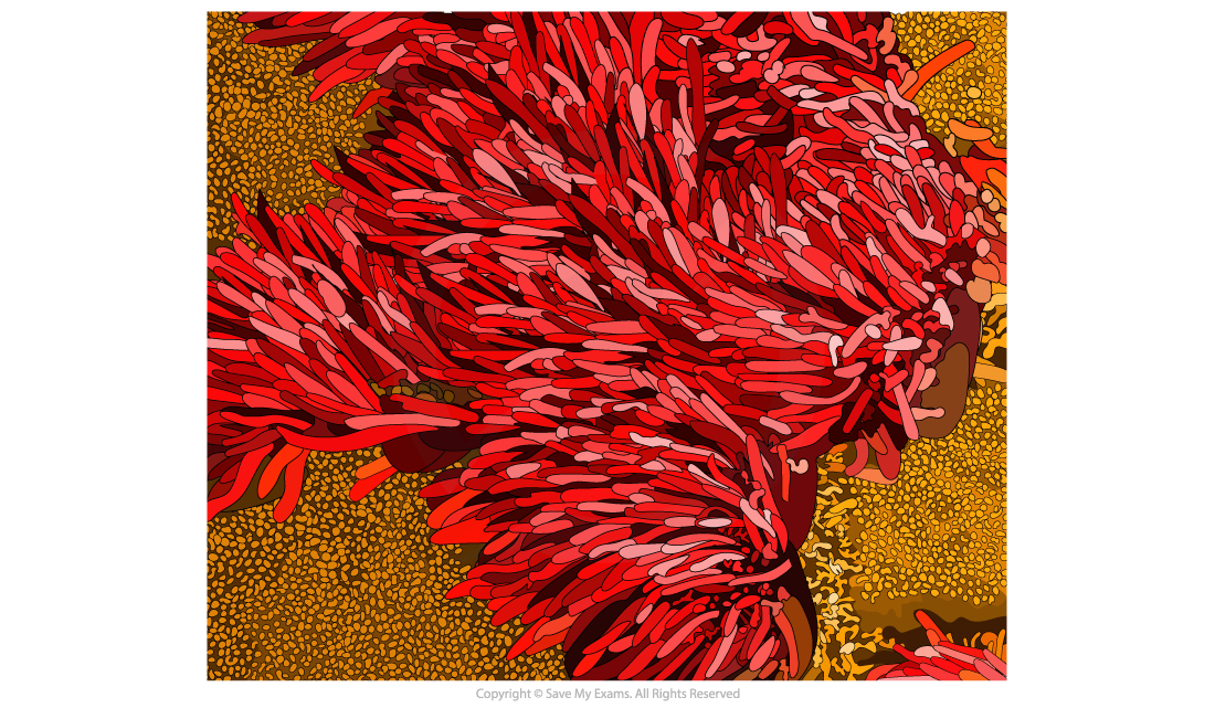
  • The density of the cilia are shown in the electron micrograph below

    • The cilia are essential for sweeping bacteria and dust-filled mucus away from the lungs and up the trachea into the back of the mouth

    • This mucus is then swallowed, with any pathogens hopefully destroyed by the acidic conditions in the stomach
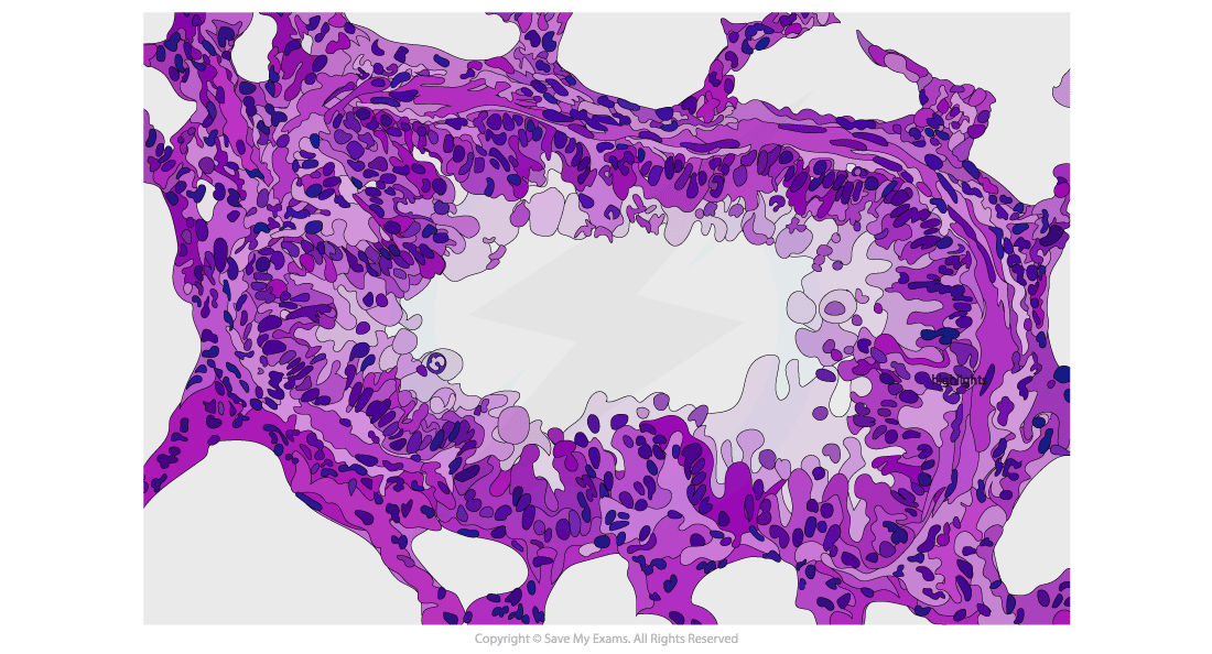
Trachea Electron Micrograph
An electron micrograph of the tissues that can be found inside the trachea

Bronchi

  • Bronchi are distinctive from the trachea because their lumen is narrower (around 8-9mm instead of 18mm

  • However, like the trachea, they are lined by ciliated epithelium

A light micrograph image showing tissue of the bronchi in cross section
A photomicrograph image showing tissue of the bronchi in cross section

Bronchioles

  • Bronchioles are approximately 1mm or less in diameter

  • Smooth muscle and cuboidal epithelium are found in their walls
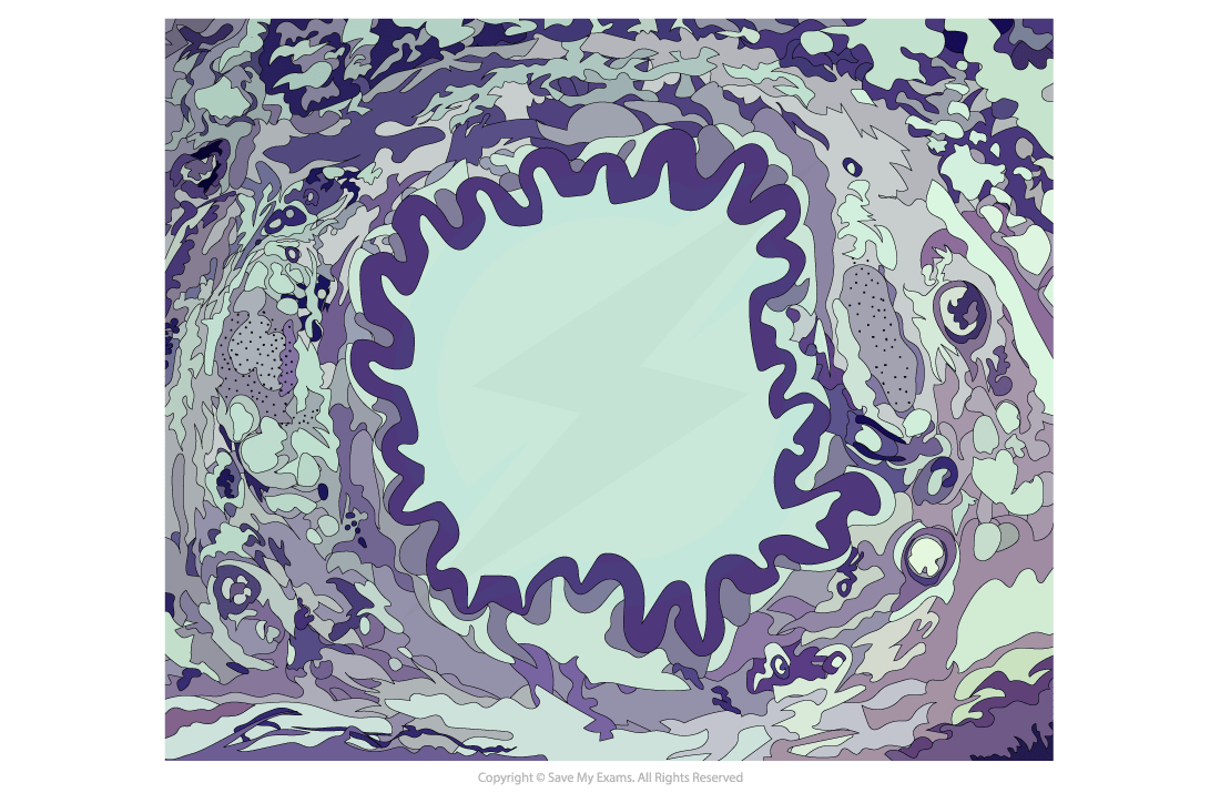
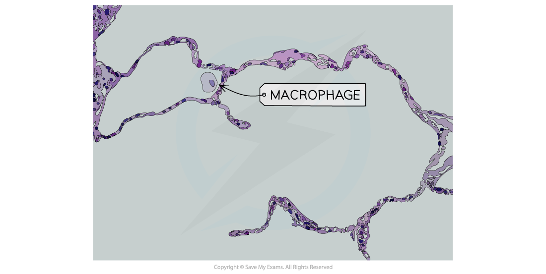
A photomicrograph showing bronchiole tissues in the gas exchange system

Alveoli

  • Alveoli have a sponge-like appearance under a microscope due to their air spaces.

  • They are surrounded by an extensive capillary network and are lined by squamous epithelium

The cells surrounding the alveolus air sac as viewed by a light microscope

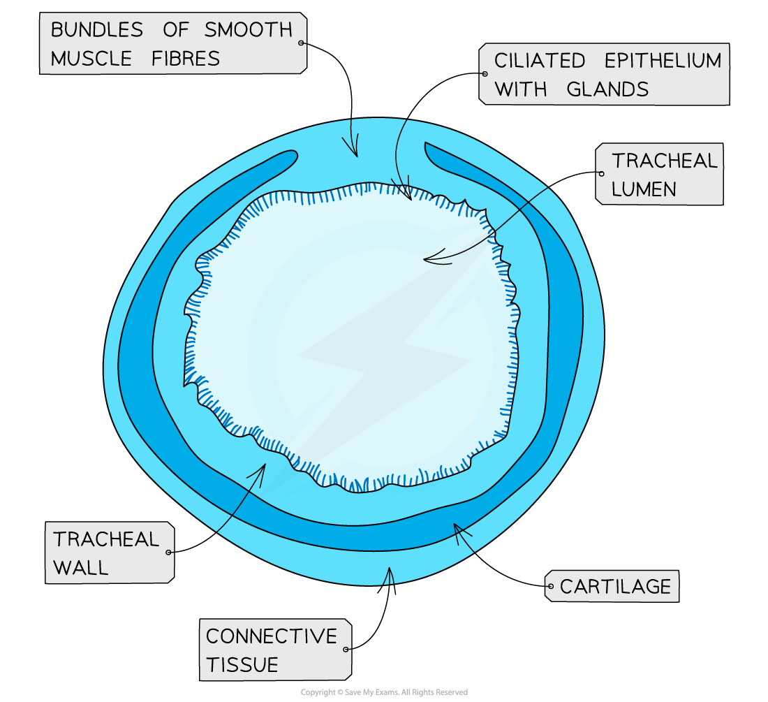
Walls of the trachea and bronchus

Trachea

  • The tracheal lumen is around 15 to 20 mm and is lined by ciliated epithelia

  • The tracheal wall is surrounded by strong and flexible cartilage which flexes during breathing

  • The smooth muscle of the trachea constricts and allows air to be expelled with more force
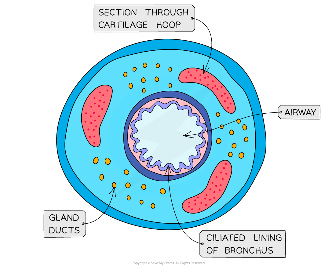
Cross-section diagram of the trachea showing labels for smooth muscle fibres, ciliated epithelium with glands, lumen, wall, cartilage, and connective tissue.
Labelled diagram of a cross section of the trachea

 Bronchus

  • Like the trachea, the bronchus is lined with ciliated epithelium

  • The cartilage hoops provide structural support to the bronchi and the gland ducts secrete mucus in the bronchial tubes

Cross-section diagram of a bronchus showing labelled sections: airway, cartilage hoop, gland ducts, ciliated lining, and surrounding tissue features.
Parts of the cross section of a bronchus. This would be the same in both bronchi

Unlock more, it's free!

Join the 100,000+ Students that ❤️ Save My Exams

the (exam) results speak for themselves:

Emma Archbold

Author: Emma Archbold

Expertise: Biology Content Creator

Prior to working at SME, Emma was a Biology teacher for 5 years. During those years she taught three different GCSE exam boards and two A-Level exam boards, gaining a wide range of teaching expertise in the subject. Emma particularly enjoys learning about ecology and conservation. Emma is passionate about making her students achieve the highest possible grades in their exams by creating amazing revision resources!

Alistair Marjot

Reviewer: Alistair Marjot

Expertise: Environmental Systems and Societies & Biology Content Creator

Alistair graduated from Oxford University with a degree in Biological Sciences. He has taught GCSE/IGCSE Biology, as well as Biology and Environmental Systems & Societies for the International Baccalaureate Diploma Programme. While teaching in Oxford, Alistair completed his MA Education as Head of Department for Environmental Systems & Societies. Alistair has continued to pursue his interests in ecology and environmental science, recently gaining an MSc in Wildlife Biology & Conservation with Edinburgh Napier University.