Practical: Microscopy (SQA National 5 Biology): Revision Note
Exam code: X807 75

Written by: Cara Head
Updated on
Examining cells using a light microscope
Many biological structures are too small to be seen by the naked eye
Light (optical) microscopes are an invaluable tool for scientists as they allow for tissues, cells and organelles to be seen and studied
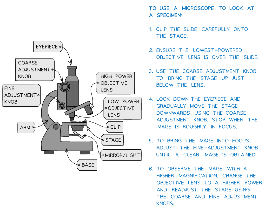
Apparatus
The key components of an optical microscope you will need to use are:
The eyepiece lens
The objective lenses
The stage
The light source
The coarse and fine focus
Other apparatus used:
Forceps
Scissors
Scalpel
Coverslip
Slides
Pipette
Stain e.g. iodine solution or methylene blue
Viewing plant tissue
Method
An ideal tissue is the onion epidermis (found between the layers of onions) because it forms a layer just one cell thick
Being a non-photosynthetic tissue, the onion epidermis is not green as it does not contain any chloroplasts
This tissue can be stained with iodine solution
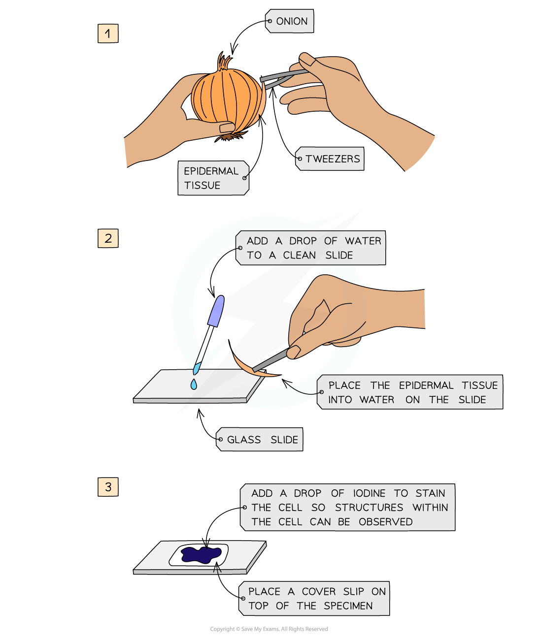
Viewing plant tissue under a light microscope
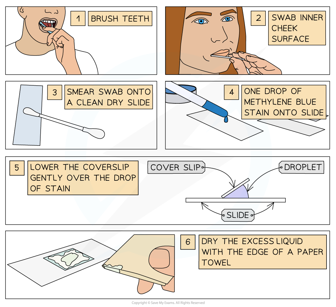
Viewing animal tissue
Method
Human cheek cells are a good choice for examination under the light microscope because they are:
plentiful
easy to obtain safely
can be obtained without an overly intrusive process
relatively unspecialised and so will display the main cell structures
Cheek cells can be stained with methylene blue solution
Extra safety precautions should be taken when using human tissue to ensure that:
the volunteer does not have a cold, cough, or throat infection that could infect someone else
all equipment is appropriately sterilised
Examiner Tips and Tricks
In addition to onion and cheek tissue, you may also examine a range of plant, animal and microbial cells using a light such as rhubarb epidermis, yeast and prepared slides of bacterial cells.

Unlock more, it's free!
Join the 100,000+ Students that ❤️ Save My Exams
the (exam) results speak for themselves:

Was this revision note helpful?