Stimulating Contraction in Striated Muscle (Cambridge (CIE) A Level Biology): Revision Note
Exam code: 9700

Stimulating contraction in striated muscle
Striated muscle contracts when it receives an impulse from a motor neurone via the neuromuscular junction
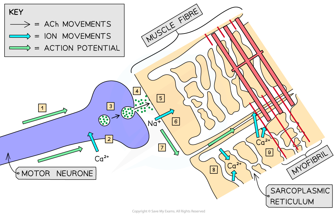
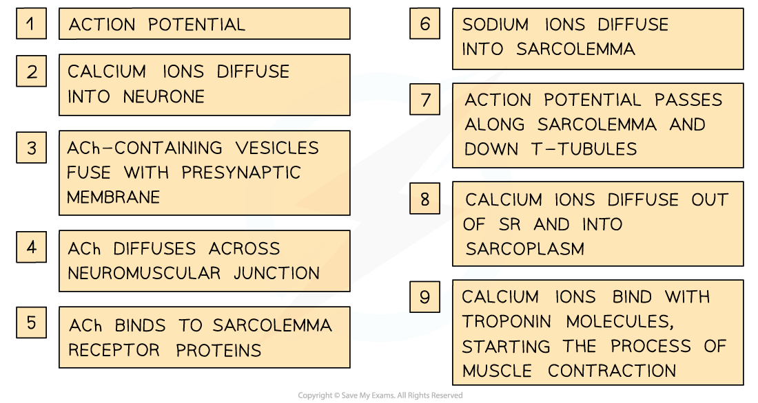
When an impulse travelling along the axon of a motor neurone arrives at the presynaptic membrane, the action potential causes calcium ions to diffuse into the neurone
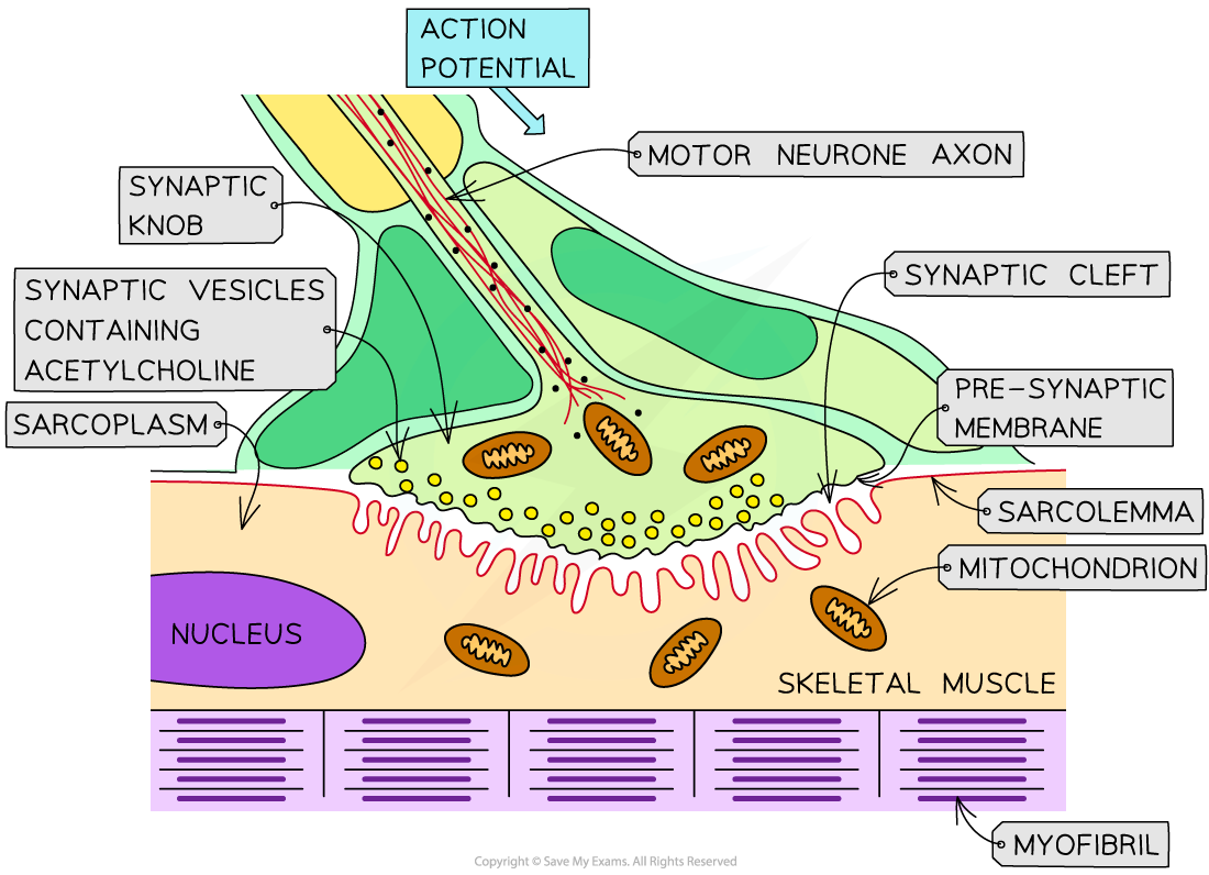
Inward diffusion of Ca2+ ions stimulates vesicles containing the neurotransmitter acetylcholine (ACh) to fuse with the presynaptic membrane
The ACh that is released diffuses across the neuromuscular junction and binds to receptor proteins on the sarcolemma (surface membrane of the muscle fibre cell)
This stimulates ion channels in the sarcolemma to open, allowing sodium ions to diffuse in
This depolarises the sarcolemma, generating an action potential that passes down the T-tubules towards the centre of the muscle fibre
These action potentials cause voltage-gated calcium ion channel proteins in the membranes of the sarcoplasmic reticulum (which lie very close to the T-tubules) to open
Calcium ions diffuse out of the sarcoplasmic reticulum (SR) and into the sarcoplasm surrounding the myofibrils
Calcium ions bind to troponin molecules, stimulating them to change shape
This causes the troponin and tropomyosin proteins to change position on the thin (actin) filaments
The myosin-binding sites are exposed on the actin molecules
The process of muscle contraction (known as the sliding filament model) can now begin
Examiner Tips and Tricks
You may have noticed that there are a lot of similarities between the events at the neuromuscular junction and those that occur at cholinergic synapses. A cholinergic synapse is between two neurones, a neuromuscular junction is between a neurone and muscle. Make sure you understand the similarities and differences so that you don’t get confused between the two.

Unlock more, it's free!
Join the 100,000+ Students that ❤️ Save My Exams
the (exam) results speak for themselves:

Was this revision note helpful?
