Ultrastructure of Striated Muscle (Cambridge (CIE) A Level Biology): Revision Note

Exam code: 9700

Phil

Written by: Phil

Reviewed by: Alistair Marjot

Updated on

Ultrastructure of striated muscle

  • Striated muscle makes up the muscles in the body that are attached to the skeleton

    • 'Striated' means it is striped/streaky in appearance

Histological slide of skeletal muscle tissue showing elongated muscle fibres in pink with multiple nuclei, and visible striations and connective tissue.
Skeletal striated muscle as seen under the microscope, after staining Image by Nephron, CC BY-SA 3.0, via Wikimedia Commons
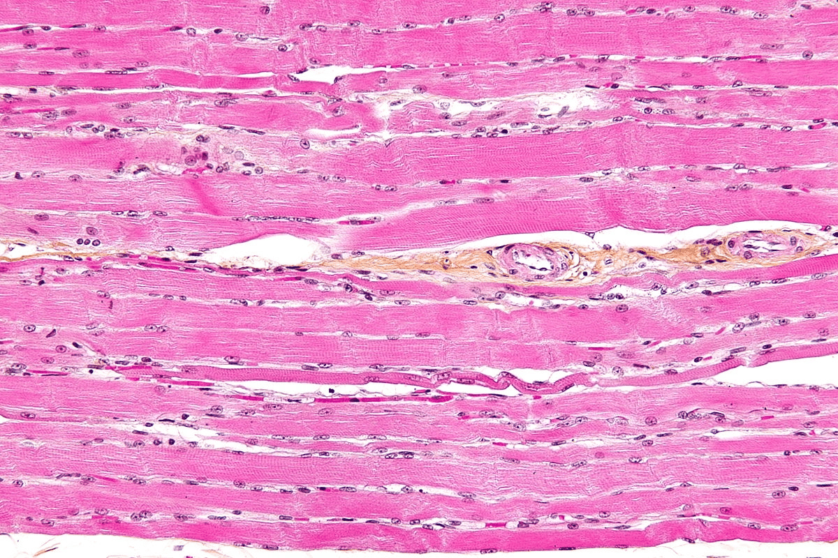
  • Striated muscle is made up of muscle fibres

  • A muscle fibre is a highly specialised cell-like unit:

    • Each muscle fibre contains an organised arrangement of contractile proteins in the cytoplasm

    • Each muscle fibre is surrounded by a cell surface membrane

    • Each muscle fibre contains many nuclei—this is why muscle fibres are not usually referred to as cells

  • The different parts of a muscle fibre have different names from the equivalent parts of a normal cell:

    • Cell surface membrane = sarcolemma

    • Cytoplasm = sarcoplasm

  • The sarcoplasm contains mitochondria and myofibrils

    • The mitochondria carry out aerobic respiration to generate the ATP required for muscle contraction

    • Myofibrils are bundles of actin and myosin filaments, which slide past each other during muscle contraction
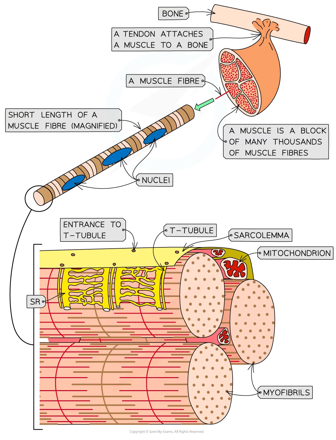
Diagram of muscle structure showing muscle fibres, tendon attachment, nuclei, sarcolemma, T-tubule, mitochondrion, sarcoplasmic reticulum, and myofibrils.
The ultrastructure of striated muscle and of a section of muscle fibre

Myofibrils

  • Myofibrils are located in the sarcoplasm

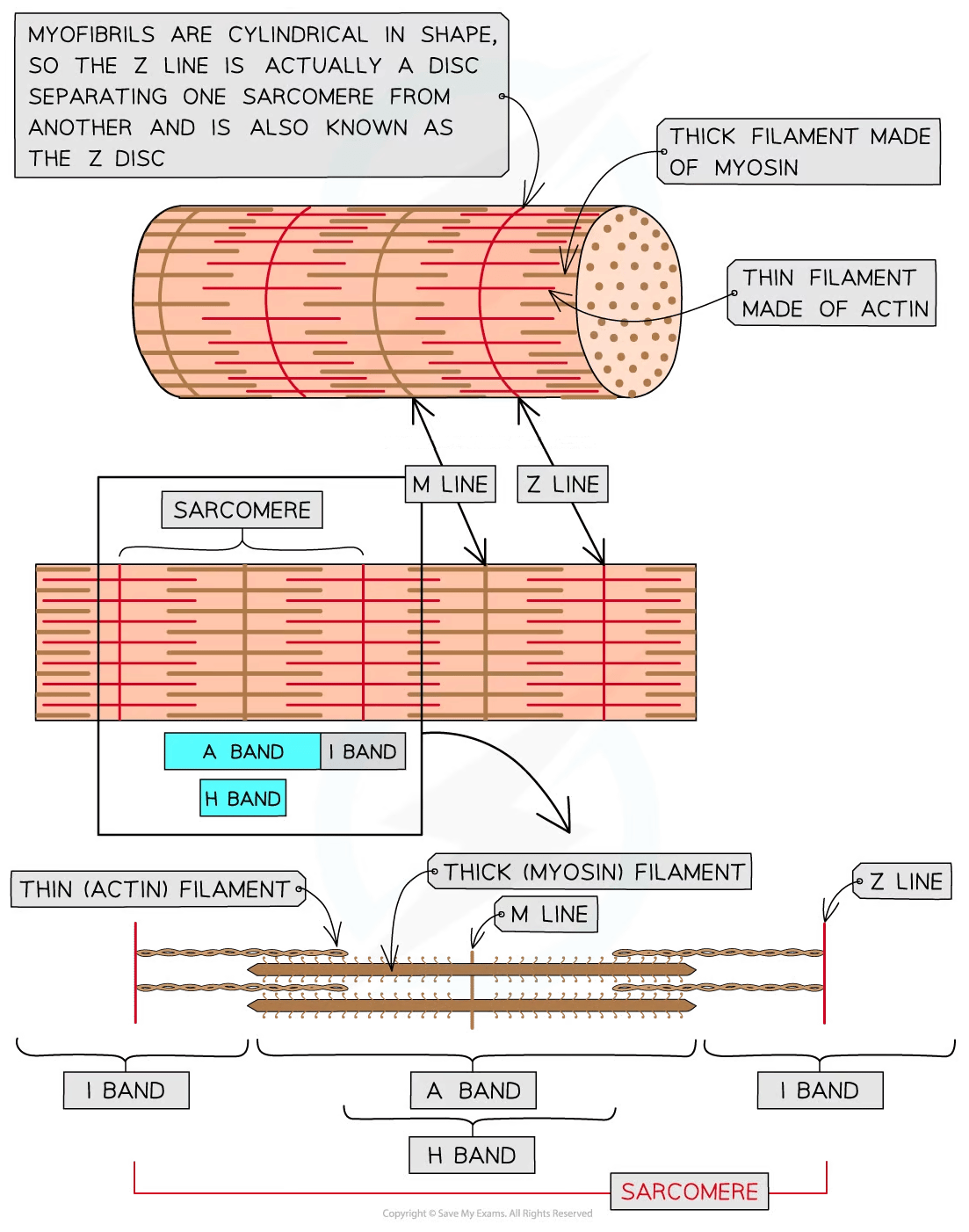
  • Each myofibril is made up of two types of protein filament:

    • Thick filaments made of myosin

    • Thin filaments made of actin

  • These two types of filament are arranged in a particular order, creating different types of band and line

Part of myofibril

Description

H band

Only thick myosin filaments present

I band

Only thin actin filaments present

A band

Contains areas where only myosin filaments are present and areas where myosin and actin filaments overlap

M Line

Attachment for myosin filaments

Z line

Attachment for actin filaments

Sarcomere

The section of myofibril between two Z lines

Diagram of a sarcomere showing myofibril structure with thick myosin filaments and thin actin filaments, including Z line, M line, A band, I band, and H band.
The structure of a myofibril

Unlock more, it's free!

Join the 100,000+ Students that ❤️ Save My Exams

the (exam) results speak for themselves:

Phil

Author: Phil

Expertise: Biology Content Creator

Phil has a BSc in Biochemistry from the University of Birmingham, followed by an MBA from Manchester Business School. He has 15 years of teaching and tutoring experience, teaching Biology in schools before becoming director of a growing tuition agency. He has also examined Biology for one of the leading UK exam boards. Phil has a particular passion for empowering students to overcome their fear of numbers in a scientific context.

Alistair Marjot

Reviewer: Alistair Marjot

Expertise: Environmental Systems and Societies & Biology Content Creator

Alistair graduated from Oxford University with a degree in Biological Sciences. He has taught GCSE/IGCSE Biology, as well as Biology and Environmental Systems & Societies for the International Baccalaureate Diploma Programme. While teaching in Oxford, Alistair completed his MA Education as Head of Department for Environmental Systems & Societies. Alistair has continued to pursue his interests in ecology and environmental science, recently gaining an MSc in Wildlife Biology & Conservation with Edinburgh Napier University.