Cells of the Blood (Cambridge (CIE) AS Biology): Revision Note
Exam code: 9700

Cells of the blood
Blood is a tissue composed of a number of important specialised cells, including:
Red blood cells
Monocytes
Neutrophils
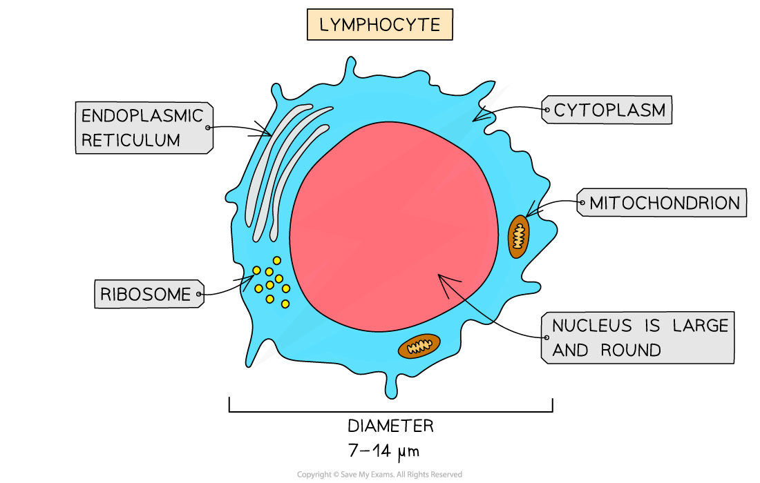
Lymphocytes
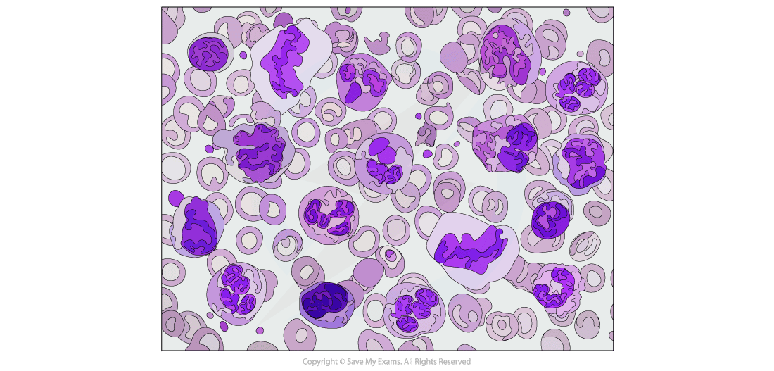
These cells all have distinguishable structures which enable them to be recognised on microscope slides, in photomicrographs and in electron micrographs
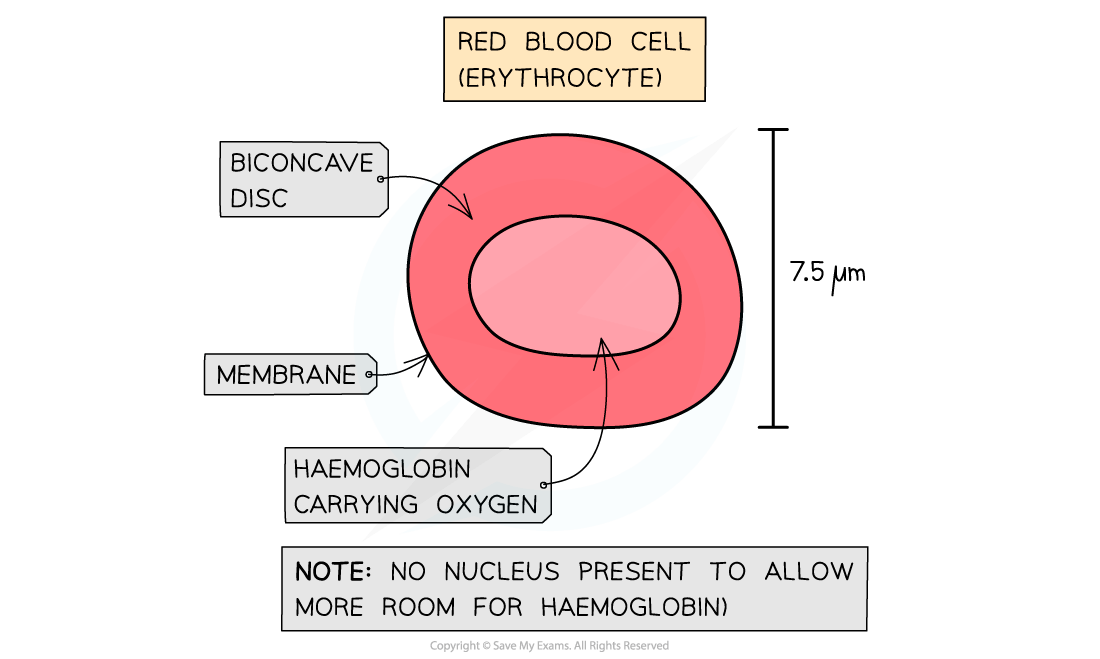
Red blood cells contain haemoglobin
This is a protein with a quaternary structure that contains haem iron groups which can bind reversibly to oxygen
Distinctive features of red blood cells when viewed under a microscope, are their distinctive biconcave disc shape (caused by their lack of nucleus)
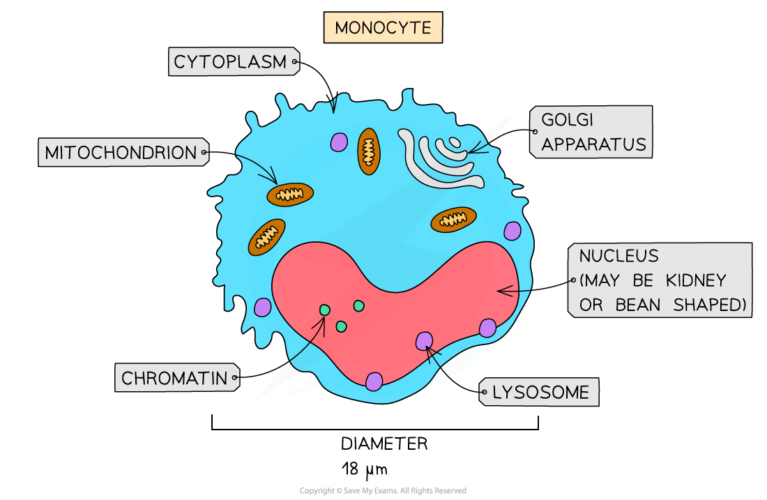
Monocytes are identifiable by their size
They are the largest of the leukocytes and have a nucleus shaped like a kidney or a bean
The nucleus of monocytes tends to appear lighter after staining than other leukocytes
The nucleus should appear a light blue colour, while the chromatin inside is distinct and fine

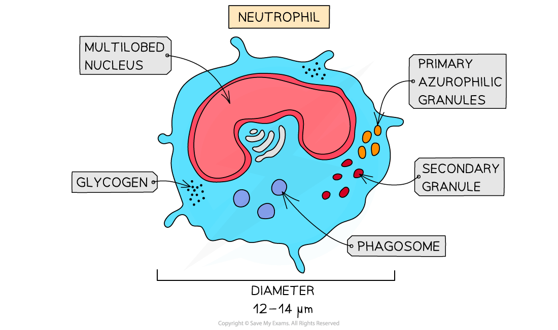
Neutrophils are distinguished by their multi-lobed nuclei
Up to 70% of all leukocytes are neutrophils
This makes them easy to spot on a micrograph
The granules of neutrophils typically stain pink or purple-blue

Lymphocytes are small leukocytes that are identifiable by their very large nuclei, which typically stains a dark colour
Lymphocytes constitute around 20-25% of all leukocytes
Lymphocytes are around the size of red blood cells
Examiner Tips and Tricks
When looking at micrographs, ensure you distinguish between the kidney-shaped nucleus of a monocyte and the multi-lobed nucleus of a neutrophil, as these can appear similar at first. As with all things, practice is key here!

Unlock more, it's free!
Join the 100,000+ Students that ❤️ Save My Exams
the (exam) results speak for themselves:

Was this revision note helpful?
