Ribosome - GCSE Biology Definition
Reviewed by: Dr Natalie Lawrence
Last updated
Key Takeaways
A ribosome is a tiny structure found in the cells of all types of organisms that builds proteins by reading the mRNA code
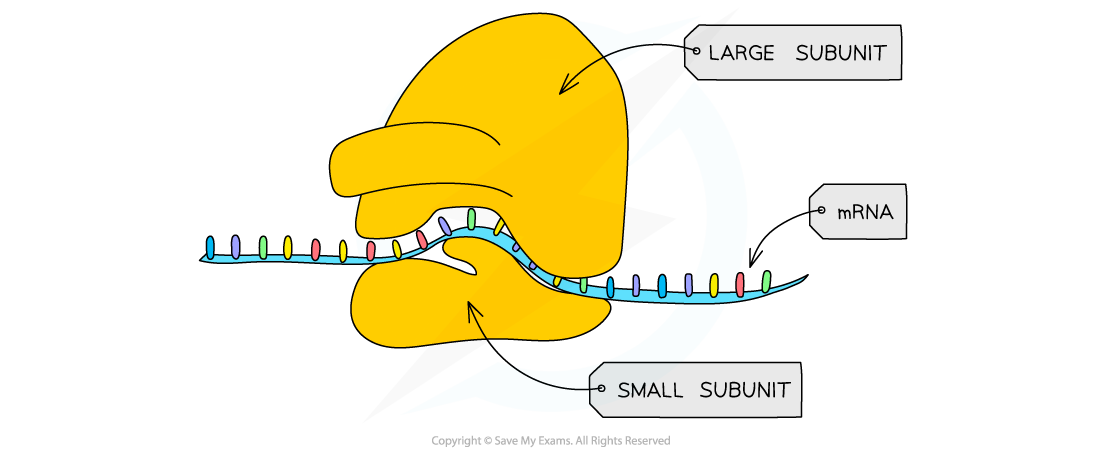
Ribosomes are made of ribosomal RNA (rRNA) and proteins, arranged into two subunits: a large subunit and a small subunit
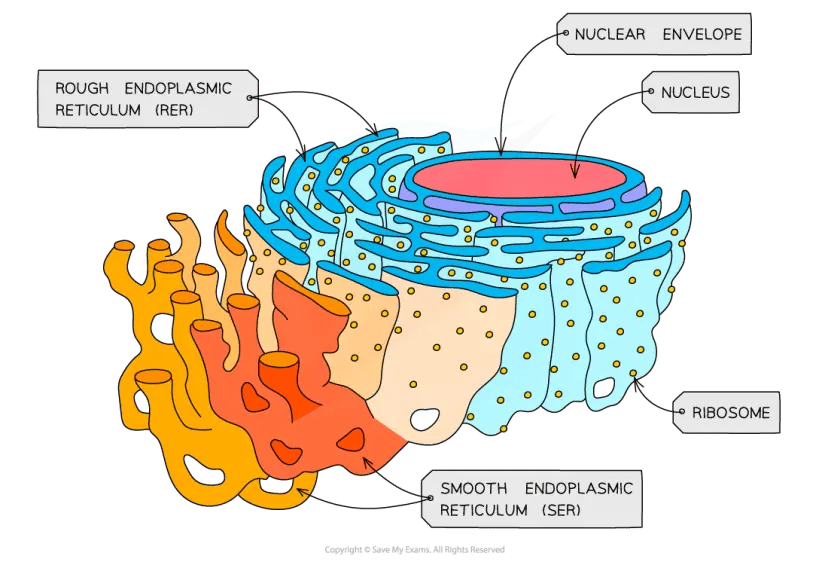
Almost every cell in your body contains ribosomes, both in the cytoplasm (free ribosomes) and attached to the rough endoplasmic reticulum (bound ribosomes)
Prokaryotic cells have smaller 70S ribosomes, while eukaryotic cells have larger 80S ribosomes
Ribosomes don't have a membrane surrounding them, so technically they aren't classified as organelles in the traditional sense
What Is a Ribosome?
A ribosome is a small organelle responsible for making proteins. You'll find ribosomes in the cytoplasm of every living cell, from bacteria to animal cells.
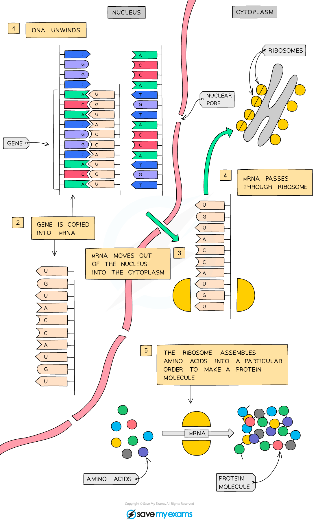
Here's what makes them so critical. DNA holds the instructions for making proteins, but DNA is far too large to leave the nucleus. Instead, the cell copies those instructions onto a smaller molecule called messenger RNA (mRNA), which travels out of the nucleus to the ribosomes. The ribosome then ‘reads’ the mRNA code and assembles the correct amino acids into a protein chain. Without ribosomes, cells couldn't produce enzymes, hormones, or structural proteins.
Both prokaryotic cells (like bacteria) and eukaryotic cells (like plant and animal cells) contain ribosomes, though there are differences in size and structure between the two.
Ribosome Structure and Subunits
Every ribosome consists of two parts: a large subunit and a small subunit. These fit together around a strand of mRNA during protein synthesis, then separate again once the job is done.
Ribosomes are made from two types of molecule: ribosomal RNA (rRNA) and proteins. The rRNA joins the amino acids together, while the proteins help stabilise the overall structure.
Component | Role |
|---|---|
Small subunit | Binds to the mRNA strand and reads the genetic code |
Large subunit | Catalyses the formation of peptide bonds between amino acids |
rRNA | Provides the enzymatic activity for protein assembly |
Proteins | Provide structural support and stability |
Unlike organelles such as mitochondria or the nucleus, ribosomes aren't enclosed by a lipid bilayer - they lack a surrounding membrane. This is why some biologists hesitate to call them true organelles, though they're still seen as key sub-cellular structures in biology.
70S and 80S Ribosomes
The "S" in 70S and 80S stands for Svedberg units: a measure of how fast a particle settles during centrifugation. It reflects size and shape rather than weight, which is why the numbers don't add up in a straightforward way.
Prokaryotic cells (bacteria) contain 70S ribosomes, made up of a 30S small subunit and a 50S large subunit. These are smaller overall.
Eukaryotic cells contain 80S ribosomes, composed of a 40S small subunit and a 60S large subunit. These are found in animal cells, plant cells, and fungi.
Ribosome Function in Protein Synthesis
Ribosomes carry out translation, the second stage of protein synthesis. During translation, the ribosome reads the mRNA code and builds a chain of amino acids that will fold into a functional protein.
The process works in three broad steps:
Initiation — the small subunit of the ribosome binds to the mRNA strand. The large subunit then joins to form the complete ribosome around the mRNA.
Elongation — the ribosome reads the mRNA in groups of three bases, called codons. Each codon specifies a particular amino acid. Transfer RNA (tRNA) molecules carry the correct amino acids to the ribosome, where their anticodon matches the mRNA codon. Two tRNA molecules sit on the ribosome at any one time, allowing a peptide bond to form between their amino acids. The ribosome then shifts along the mRNA to read the next codon.
Termination — when the ribosome reaches a stop codon on the mRNA, translation ends. The completed amino acid chain is released and folds into its final protein shape.
Once released, the protein folds into a unique three-dimensional shape that determines its function. It might become an enzyme that speeds up chemical reactions, a hormone that carries messages around the body, or a structural protein like collagen that strengthens tissues.
“I always suggest my students find a good video of the translation process on YouTube or elsewhere, because static images make it very difficult to appreciate how it happens. It really helps to see the tRNA molecules entering the ribosome with their amino acids and leaving empty as the polypeptide grows.”
– Natalie Lawrence, Biology Tutor.
Save My Exams covers the full process of protein synthesis, including transcription and translation, with clear diagrams and examiner tips. Our revision notes break down each stage and explain how the ribosome reads mRNA codons to assemble amino acids in sequence. Explore the topic further with Protein Synthesis - Edexcel GCSE Biology Revision Notes or you can find the notes specific to your course.
Free and Bound Ribosomes
Not all ribosomes do the same job, and their location within the cell gives a clue about what they're producing.
Free ribosomes float in the cytoplasm, unattached to any membrane. They tend to produce proteins that function inside the cell itself, such as enzymes used in metabolic reactions or proteins needed for cell growth.
Bound ribosomes are attached to the surface of the rough endoplasmic reticulum (rough ER), which is what gives it its "rough" appearance under an electron microscope. These ribosomes produce proteins destined to be exported out of the cell, inserted into the cell membrane, or packaged into vesicles. Digestive enzymes and antibodies are good examples.
Ribosomes and Antibiotics
The structural difference between prokaryotic 70S ribosomes and eukaryotic 80S ribosomes has a practical application that affects millions of people.
Many antibiotics work by targeting the 70S ribosomes in bacteria. Tetracycline, for instance, blocks tRNA from attaching to the bacterial ribosome, stopping protein synthesis. Erythromycin prevents the ribosome from moving along the mRNA strand. In both cases, the bacterium can't make the proteins it needs and dies.
Because human cells use 80S ribosomes with a different structure, these antibiotics don't interfere with our own protein production. But there's a catch: mitochondria inside our cells also contain 70S ribosomes (a remnant of their evolutionary origin as free-living bacteria). At very high doses, some antibiotics can affect mitochondrial function.
If you're building your understanding of cell structures and how sub-cellular components like ribosomes work together, Save My Exams revision notes cover eukaryotic and prokaryotic cells side by side, with labelled diagrams and exam-style practice. See our AQA GCSE notes on Eukaryotes & Prokaryotes or find the notes tailored to your specific course.
Frequently Asked Questions
What is the difference between a ribosome and a mitochondrion?
Ribosomes and mitochondria do different jobs. A ribosome builds proteins by reading mRNA instructions. A mitochondrion generates energy for the cell through aerobic respiration. Structurally, mitochondria are much larger, enclosed by a double membrane, and contain their own DNA. Ribosomes are smaller, have no membrane, and are made of rRNA and protein.
Are ribosomes considered organelles?
Strictly speaking, ribosomes aren't membrane-bound, which is the traditional requirement for an organelle. Most biology courses still list them as sub-cellular structures or sometimes as non-membrane-bound organelles.
How many ribosomes does a cell have?
A single human cell can contain millions of ribosomes. Cells that produce large quantities of protein, such as liver cells or cells lining the digestive system, tend to have even more. Bacterial cells are smaller but can still contain tens of thousands.
Where are ribosomes made in the cell?
In eukaryotic cells, the components of ribosomes are produced in the nucleolus, a dense region within the nucleus. The ribosomal subunits are then exported through nuclear pores into the cytoplasm, where they come together around an mRNA strand to begin translation.
Examiner-written GCSE Biology revision resources that improve your grades 2x
- Written by expert teachers and examiners
- Aligned to exam specifications
- Everything you need to know, and nothing you don’t
Was this glossary entry helpful?

Share this article

 written revision resources that improve your