Scale Bar - GCSE Biology Definition
Reviewed by: Dr Natalie Lawrence
Last updated
Key Takeaways
A scale bar is a line on a diagram, micrograph, or map that represents a known real-world distance
In biology, scale bars show the actual size of cells and structures viewed under a microscope, typically measured in micrometres (µm) or nanometres (nm)
You can calculate actual size using the formula: actual size = image size ÷ magnification
Scale bars are more reliable than magnification statements alone because they stay accurate even when an image is resized
Converting between units (mm, µm, nm) is a key skill when working with scale bars in biology
What Is a Scale Bar?
A scale bar is a straight line printed on an image or diagram that represents a specific real-world measurement. Think of it as a built-in ruler measurement. It tells you exactly how large or small the real object is compared to the images you're looking at on paper or on a screen.
Scale bars appear across science, geography, and engineering. You'll find them on microscope images, maps, architectural plans, and technical drawings. They all do the same job: connect what you see in the image to a real measurement.
Scale Bars in Biology
In biology, scale bars are most common on micrographs (photographs taken through a microscope). Cells and subcellular structures are far too small to see with the naked eye, so biologists rely on microscopes to magnify them. A scale bar on a micrograph might show that a short line represents 10 µm or 50 nm, depending on the level of magnification used.
This matters because biological specimens vary hugely in size. A typical animal cell measures between 10 and 100 µm, while a bacterium might be just 1 µm across. That's a difference of several orders of magnitude. Without a scale bar, there's no way to tell how big something actually is from the image alone.
Scale Bars on Maps
On a map, a scale bar works the same way but at a much larger scale. A line might represent 1 km or 10 miles, for instance. Map scale bars let you estimate real distances between locations without needing to know the exact ratio of the map. If you shrink or enlarge the map, the scale bar changes with it, so it always stays accurate.
How to Draw a Scale Bar
Drawing a scale bar on a biological diagram is a practical skill that comes up regularly. Here's the process:
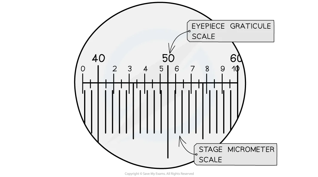
Measure the specimen using the eyepiece graticule or by calculating its actual size from the magnification
Choose a round number that fits neatly on your drawing (e.g. 10 µm, 50 µm, 100 µm)
Calculate the length the bar should be on your drawing using: image size = actual size × magnification
Draw a straight line of that length, using a ruler
Label it clearly with the measurement it represents (e.g. "50 µm")
Place the scale bar near the bottom of your drawing, away from the main image. It should sit horizontally, with flat ends (not arrows), and always include the unit.
Worked Example
Imagine you're drawing a cell observed at ×400 magnification. You know the cell's actual diameter is 50 µm.
To find how long the scale bar should be on your drawing, multiply: 50 × 400 = 20,000 µm, which is 20 mm.
So a 20 mm line on your drawing represents 50 µm. You'd label it "50 µm."
If you want a tidier scale bar, you could represent 10 µm instead: 10 × 400 = 4,000 µm = 4 mm. A 4 mm line labelled "10 µm" works just as well and keeps the drawing uncluttered.
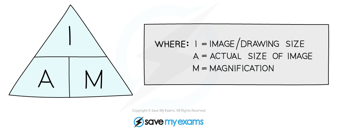
Scale Bar Calculations
Three quantities link together when working with scale bars and microscopy:
Quantity | What it means | Unit |
|---|---|---|
Image size | The measured length on the drawing or photograph | Usually mm |
Actual size | The real size of the specimen | Usually µm or nm |
Magnification | How many times the image has been enlarged | No unit (written as ×400, ×1000, etc.) |
The magnification equation ties them together:
Magnification = image size ÷ actual size
Actual size = image size ÷ magnification
Image size = actual size × magnification
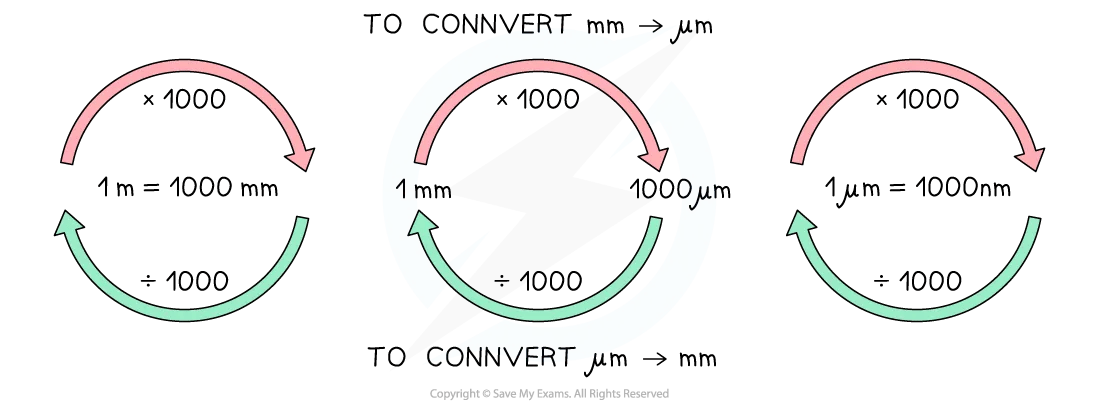
Unit conversions are where most errors creep in. Both measurements must be in the same unit before you calculate. The key conversions:
1 mm = 1,000 µm
1 µm = 1,000 nm
Therefore:
To go from µm to mm, divide by 1,000
To go from mm to µm, multiply by 1,000
Common Mistakes With Scale Bars
Even confident students trip up on scale bars. Watch out for these:
Forgetting to convert units before calculating. If your image is measured in mm but the actual size is given in µm, the answer will be wrong by a factor of 1,000
Leaving the scale bar unlabelled. A line without a measurement and unit is meaningless
Confusing magnification with resolution. Magnification makes things look bigger; resolution determines how much detail you can actually see. A blurry image at ×10,000 hasn't gained any useful detail over a clear image at ×1,000
Assuming the scale bar becomes inaccurate after resizing. If someone photocopies or digitally resizes an image, a magnification statement (e.g. "×400") becomes inaccurate. The scale bar, however, scales with the image and remains reliable
“Scale bar calculations can seem tricky at first. I always encourage my students to practice them a bit, and they soon realise that they’re quite simple.”
– Natalie Lawrence, Biology Tutor.
Save My Exams' revision notes for AQA GCSE Microscopy cover magnification calculations, unit conversions, and the equation triangle in detail, written by experienced examiners. We have specific notes for whatever course you are doing, too.
Frequently Asked Questions
How do you calculate actual size from a scale bar?
Measure the scale bar on the image (in mm), then measure the structure you're interested in (in mm). Divide the structure's measured length by the scale bar's measured length, and multiply by the distance the scale bar represents. For example, if a scale bar labelled "10 µm" measures 5 mm on the image, and your cell measures 15 mm, the actual cell size is (15 ÷ 5) × 10 = 30 µm.
What units are used on a scale bar in biology?
Micrometres (µm) for cells and large subcellular structures, and nanometres (nm) for smaller structures like ribosomes or viruses. Light microscope images typically use µm, while electron micrographs often use nm because they reveal much finer detail.
Why is a scale bar better than just stating the magnification?
Magnification statements only hold true at the original image size. The moment someone prints, projects, or resizes the image, the stated magnification no longer matches reality. A scale bar physically changes size alongside the image, so the ratio between the bar and the specimen stays constant. It's a more trustworthy reference.
How do you convert between millimetres, micrometres, and nanometres?
Multiply by 1,000 to move to a smaller unit: 1 mm = 1,000 µm, and 1 µm = 1,000 nm. Divide by 1,000 to move to a larger unit. A quick check: if your answer has jumped by a factor of a million, you've probably skipped a step or gone the wrong way.
Examiner-written GCSE Biology revision resources that improve your grades 2x
- Written by expert teachers and examiners
- Aligned to exam specifications
- Everything you need to know, and nothing you don’t

Was this glossary entry helpful?

Share this article

 written revision resources that improve your