The Fluid Mosaic Model (Cambridge (CIE) AS Biology): Revision Note
Exam code: 9700

The fluid mosaic model of membranes
Membranes are vital structures found in all cells
The cell surface membrane creates an enclosed space separating the internal cell environment from the external environment
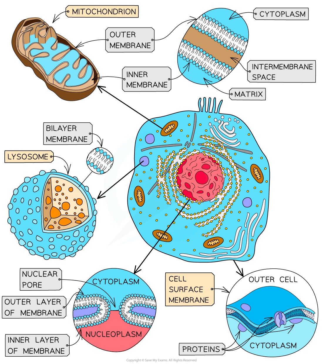
Intracellular membranes form compartments within the cell such as the nucleus, mitochondria and RER
Membranes do not only separate different areas but also control the exchange of materials across them, as well as acting as an interface for communication
Membranes are selectively permeable
Substances can cross membranes by diffusion, osmosis and active transport
Cellular membranes are formed from a bilayer of phospholipids which is roughly 7nm (7 × 10⁻⁹ metres) wide and therefore just visible under an electron microscope at very high magnifications
The fluid mosaic model of the membrane was first outlined in 1972 and it explains how biological molecules are arranged to form cell membranes
The fluid mosaic model also helps to explain:
Passive and active movement between cells and their surroundings
Cell-to-cell interactions
Cell signalling
Phospholipids
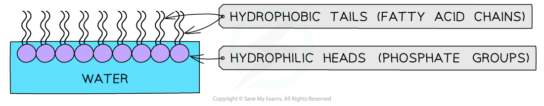
Phospholipids structurally contain two distinct regions: a polar head and two nonpolar tails
The phosphate head of a phospholipid is polar (hydrophilic) and therefore soluble in water
The lipid tail is non-polar (hydrophobic) and insoluble in water
If phospholipids are spread over the surface of water they form a single layer with the hydrophilic phosphate heads in the water and the hydrophobic fatty acid tails sticking up away from the water
This is called a phospholipid monolayer
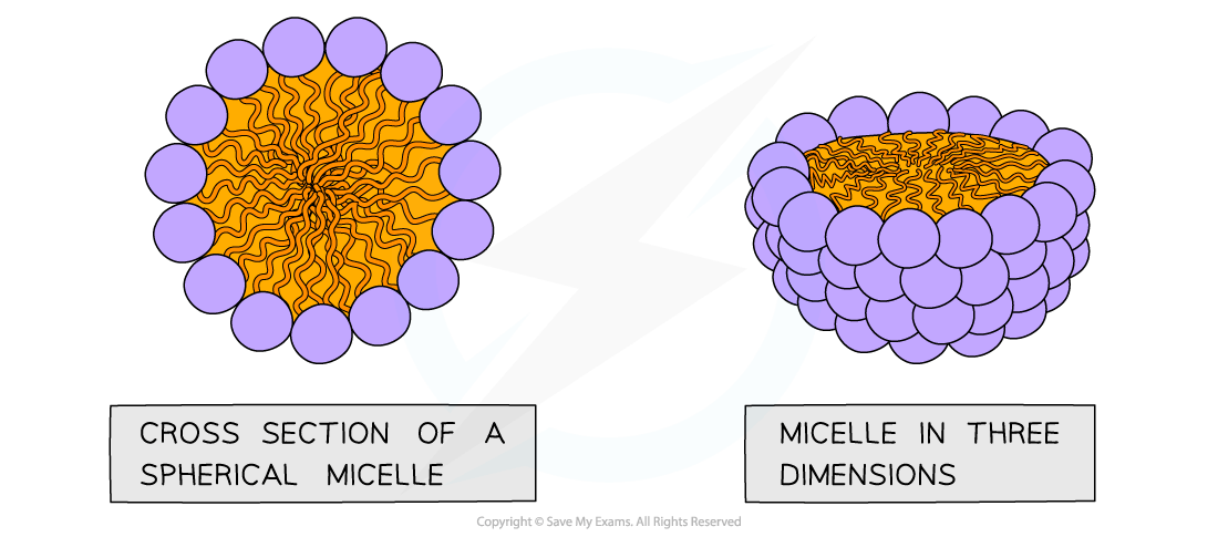
If phospholipids are mixed/shaken with water they form spheres
With the hydrophilic phosphate heads facing out towards the water, and
The hydrophobic fatty acid tails facing in towards each other
This is called a micelle
Alternatively, two-layered structures may form in sheets
These are called phospholipid bilayers – this is the basic structure of the cell membrane
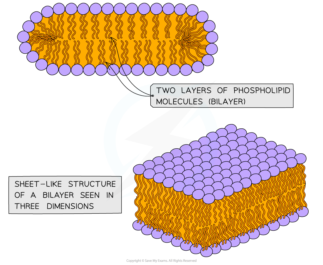
Phospholipid bilayers can form compartments – the bilayer forming the cell surface membrane establishing the boundary of each cell
Internally, membrane-bound compartments formed from phospholipid bilayers provide the basic structure of organelles
This allows for specialisation of processes within the cell
An example of a membrane-bound organelle is the lysosome (found in animal cells)
Each lysosome contains many hydrolytic enzymes that can break down many different kinds of biomolecule
These enzymes need to be kept compartmentalised otherwise they would break down most of the cellular components
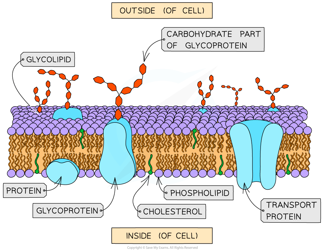
Structure of membranes
The phospholipid bilayers that make up cell membranes also contain proteins
The proteins can either be intrinsic (or integral) or extrinsic (peripheral)
Intrinsic proteins are embedded in the membrane with their arrangement determined by their hydrophilic and hydrophobic regions
Extrinsic proteins are found on the outer or inner surfaces of the membrane
The fluid mosaic model describes cell membranes as ‘fluid’ because:
The phospholipids and proteins can move around via diffusion
The phospholipids mainly move sideways, within their own layers
The many different types of protein that are interspersed throughout the bilayer move about within it (a bit like icebergs in the sea) although some may be fixed in position
The fluid mosaic model describes cell membranes as ‘mosaics’ because:
The scattered pattern produced by the proteins within the phospholipid bilayer looks somewhat like a mosaic when viewed from above
Examiner Tips and Tricks
You must know how to draw and label the fluid mosaic model, as well as ensure that you can describe why it is called the fluid mosaic model.

Unlock more, it's free!
Join the 100,000+ Students that ❤️ Save My Exams
the (exam) results speak for themselves:

Was this revision note helpful?
