The Stages of Mitosis (Cambridge (CIE) AS Biology): Revision Note
Exam code: 9700

Mitosis: the stages
Mitosis is the process of nuclear division by which two genetically identical daughter nuclei are produced that are also genetically identical to the parent cell nucleus (they have the same number of chromosomes as the parent cell)
Although mitosis is actually one continuous process, it can be divided into four main stages
These stages are:
Prophase
Metaphase
Anaphase
Telophase
Most organisms contain many chromosomes in the nuclei of their cells (e.g. humans have 46) but the diagrams below show mitosis of an animal cell with only four chromosomes, for the sake of simplicity
The different colours of the chromosomes are just to show that half are from the female parent and half from the male parent
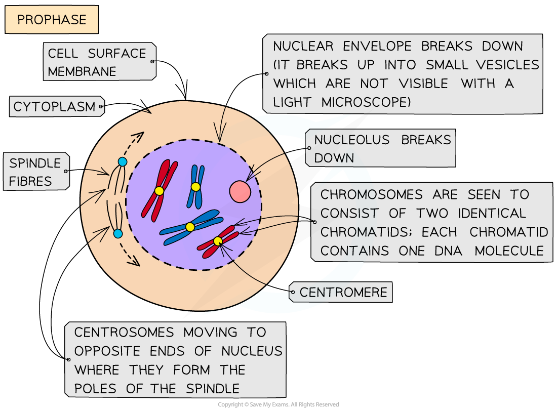
Prophase
Chromosomes condense (and are now visible when stained)
The chromosomes consist of two identical chromatids called sister chromatids
Each sister chromatid contains one DNA molecule
They are joined together at the centromere
The two centrosomes (replicated in the G2 phase just before prophase) move towards opposite poles (opposite ends of the nucleus)
Spindle fibres (protein microtubules) begin to emerge from the centrosomes (consists of two centrioles in animal cells)
The nuclear envelope (nuclear membrane) breaks down into small vesicles
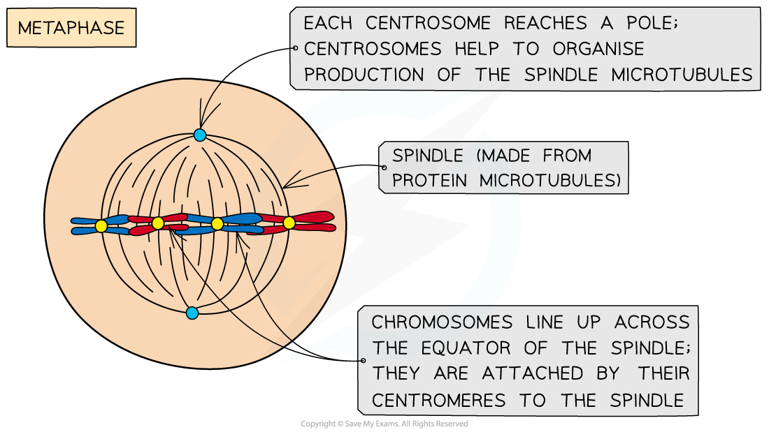
Metaphase
Centrosomes reach opposite poles
Spindle fibres (protein microtubules) continue to extend from centrosomes
Chromosomes line up at the equator of the spindle (also known as the metaphase plate) so they are equidistant to the two centrosome poles
Spindle fibres (protein microtubules) reach the chromosomes and attach to the centromeres
Each sister chromatid is attached to a spindle fibre originating from opposite poles
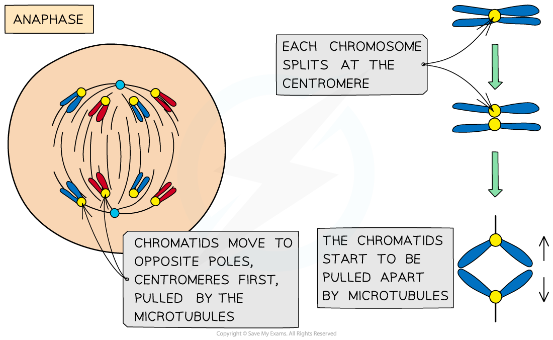
Anaphase
The sister chromatids separate at the centromere (the centromere divides in two)
Spindle fibres (protein microtubules) begin to shorten
The separated sister chromatids (now called chromosomes) are pulled to opposite poles by the spindle fibres (protein microtubules)
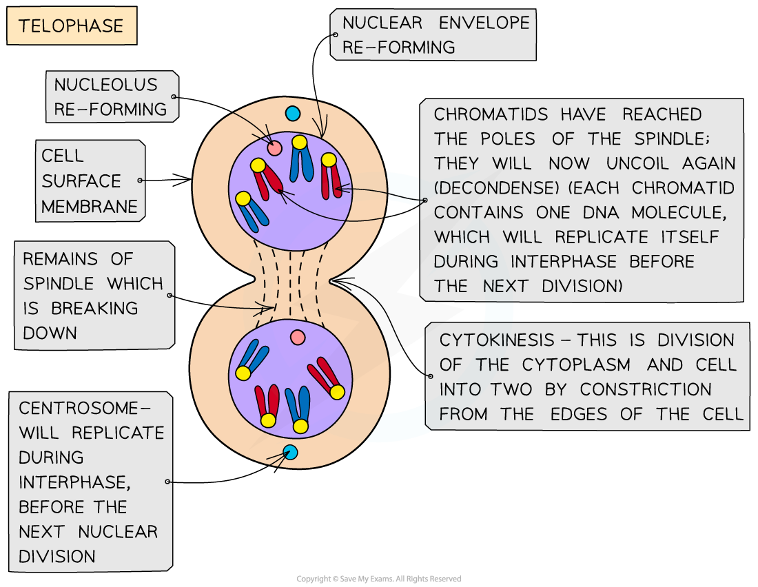
Telophase
Chromosomes arrive at opposite poles and begin to decondense
Nuclear envelopes (nuclear membranes) begin to reform around each set of chromosomes
The spindle fibres break down
Examiner Tips and Tricks
Make sure you learn the four stages of mitosis and what is happening to the DNA molecules (one chromatid contains one DNA molecule) at each stage.
Learn ‘PMAT’ (prophase, metaphase, anaphase, telophase) to help you remember the order of the stages!
After interphase but before the parent cell undergoes mitosis, the human parent cell nucleus actually contains 92 DNA molecules! This is because during interphase (S phase), the 46 DNA molecules in the parent cell have replicated to form sister chromatids. As human cells have a diploid number of 46 this replication results in 92 molecules.
This ensures the two daughter cells will be diploid (have 46 chromosomes each) when mitosis occurs. Remember to read the questions carefully as only human diploid cells have 46 chromosomes so if the question refers to another organism, its diploid number will be different.

Unlock more, it's free!
Join the 100,000+ Students that ❤️ Save My Exams
the (exam) results speak for themselves:

Was this revision note helpful?
Build on this topic
