Structure of the Heart (Cambridge (CIE) AS Biology): Revision Note
Exam code: 9700
Mammalian heart structure
Heart structure
The human heart has a mass of around 300g and is roughly the size of a closed fist
The heart is a hollow, muscular organ located in the chest cavity
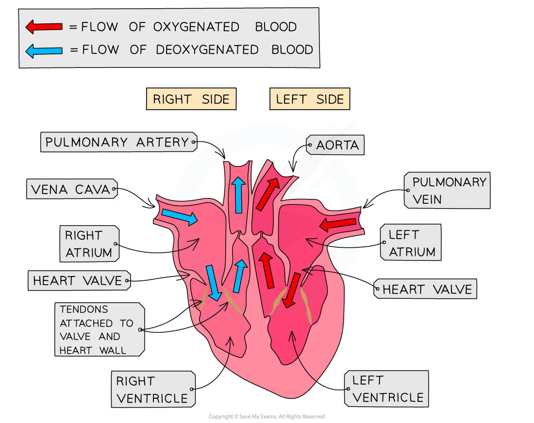
The heart is divided into four chambers
The two top chambers are atria and the bottom two chambers are ventricles
The left and right sides of the heart are separated by a wall of muscular tissue, called the septum
The septum is very important for ensuring blood doesn’t mix between the left and right sides of the heart
Valves in the heart
Valves in the heart:
Open when the pressure of blood behind them is greater than the pressure in front of them
Close when the pressure of blood in front of them is greater than the pressure behind them
Valves are important for keeping blood flowing forward in the right direction and stopping it flowing backwards
They are also important for maintaining the correct pressure in the chambers of the heart
The right atrium and right ventricle are separated by the atrioventricular valve
This is also known as the tricuspid valve
The right ventricle and the pulmonary artery are separated by the pulmonary valve
The left atrium and left ventricle are separated by the mitral valve
This is also known as the bicuspid valve
The left ventricle and aorta are separated by the aortic valve
There are two blood vessels bringing blood to the heart:
Vena cava
Pulmonary vein
There are two blood vessels taking blood away from the heart:
Pulmonary artery
Aorta
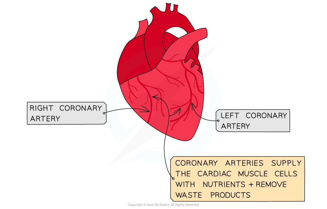
Coronary arteries
The heart is a muscle and so requires its own blood supply for aerobic respiration
The heart receives blood through arteries on its surface, called coronary arteries
It’s important that these arteries remain clear of plaques, as this could lead to angina or a heart attack (myocardial infarction)
Examiner Tips and Tricks
When looking at the heart, remember the right side of the heart will appear on the page as being on the left. This is because the heart is labelled as if it were in your body and flipped around.

Unlock more, it's free!
Join the 100,000+ Students that ❤️ Save My Exams
the (exam) results speak for themselves:

Was this revision note helpful?
