Key Takeaways
A relay neurone is a nerve cell found in the central nervous system (brain and spinal cord) that connects sensory neurones to motor neurones
Relay neurones are short, with a small cell body and many branching dendrites
They play a key role in reflex arcs, rapidly passing electrical impulses from sensory neurones to motor neurones
Signals pass between neurones at junctions called synapses
What Is a Relay Neurone?
A relay neurone is a type of nerve cell that connects sensory neurones and motor neurones. Relay neurones are found exclusively within the central nervous system, which means they're located in the brain and spinal cord.
Sensory neurones deliver electrical impulses from receptors (like pain sensors in your skin) to the CNS. The relay neurone receives that signal and passes it on to a motor neurone, which then carries the impulse to an effector, such as a muscle or gland.
You might also see relay neurones referred to as intermediate or interneurons. That's the term used more often in academic research and university-level textbooks. Both names describe the same cell doing the same job.
Relay Neurone Structure
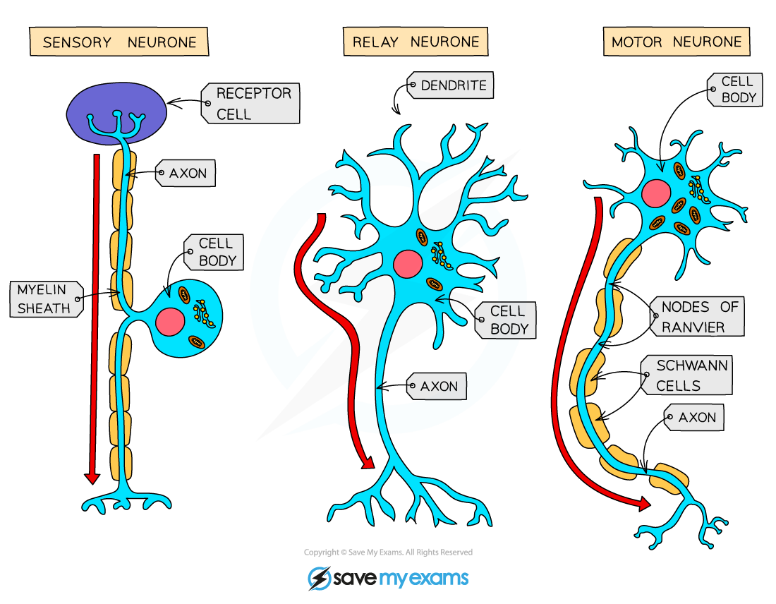
Relay neurones look quite different from sensory and motor neurones. Their structure reflects their role as connectors within the CNS.
A relay neurone has a small cell body typically located at one end, with many dendrites branching off it. These dendrites allow it to receive signals from multiple other neurones at once. The axon is short because the relay neurone only needs to transmit signals across small distances within the spinal cord or brain.
Unlike sensory and motor neurones, relay neurones have little myelin sheath. Their compact size means impulses travel short distances quickly without needing the insulation that longer neurones rely on.
What Does the Relay Neurone Do?
The relay neurone's main function is to pass electrical impulses between sensory and motor neurones during a nervous response. This is especially clear in reflex arcs, where speed is critical.
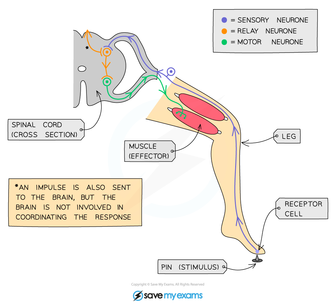
Here's how a reflex arc works, step by step:
A stimulus (e.g. stepping on a pin) is detected by a receptor in the skin
A sensory neurone sends an electrical impulse to the spinal cord
The impulse passes to a relay neurone inside the spinal cord
The relay neurone transmits the impulse to a motor neurone via a synapse
The motor neurone carries the impulse to an effector (a muscle)
The muscle contracts, pulling the foot away from the pin
This entire sequence happens in a fraction of a second. Within the brain, relay neurones form complex networks that help process and coordinate information before a response is sent out. Reflex actions bypass the conscious brain entirely, which is why you pull your hand away from a hot surface before your brain even receives the signal.
“Relay neurones and the many different synaptic connections they can make are what allow the nervous system to encode complex information. With neurones just carrying action potentials long-distances, as in motor neurones, the types of information the system could carry would be limited.”
– Natalie Lawrence, Biology Tutor.
Sensory, Relay and Motor Neurones Compared
The three types of neurone in a reflex arc each have distinct structures suited to their role. The table below summarises the main differences.
Feature | Sensory Neurone | Relay Neurone | Motor Neurone |
|---|---|---|---|
Location | Connects receptors to the CNS | Found only in the CNS | Connects the CNS to effectors |
Length | Long | Short | Long |
Cell body position | Branches off the middle of the axon | At one end, with many dendrites | At one end, with many dendrites |
Direction of impulse | From receptor to CNS | Within the CNS | From CNS to effector |
Function | Carries sensory information to the brain or spinal cord | Connects sensory and motor neurones | Carries motor commands to muscles or glands |
One thing worth noting is that the peripheral nervous system contains sensory and motor neurones, but relay neurones are found exclusively in the CNS. This distinction matters because it explains why damage to the spinal cord can disrupt reflex responses even when the sensory and motor neurones themselves are intact.
If you're revising the nervous system and want to see how these neurones work together, Save My Exams has detailed revision notes on The Reflex Arc for AQA GCSE – as well as other courses – that walk through the pathway with labelled diagrams, written by experienced biology teachers and examiners.
Frequently Asked Questions
Where are relay neurones found in the body?
Relay neurones are found exclusively in the central nervous system, meaning the brain and the spinal cord. They aren't present in the peripheral nervous system. Their position within the CNS allows them to act as the connection point between incoming sensory signals and outgoing motor commands.
What is the difference between a relay neurone and an interneuron?
There isn't one. "Relay neurone" and "interneuron" are two names for the same cell. "Relay neurone" is the term you'll encounter most often in school biology.
Why are relay neurones important in reflex arcs?
Relay neurones connect sensory neurones to motor neurones within the spinal cord, allowing reflex responses to happen without involving the conscious brain. This makes reflexes automatic and extremely fast, which helps protect the body from harm.
How do synapses connect relay neurones to other neurones?
Synapses are tiny gaps between neurones. When an electrical impulse reaches the end of one neurone, it triggers the release of chemical neurotransmitters into the gap. These chemicals diffuse across and bind to receptors on the next neurone, generating a new electrical impulse. This chemical-to-electrical conversion is how signals pass between relay neurones and their neighbouring sensory or motor neurones.
Examiner-written GCSE Biology revision resources that improve your grades 2x
- Written by expert teachers and examiners
- Aligned to exam specifications
- Everything you need to know, and nothing you don’t
Was this glossary entry helpful?


Share this article
 written revision resources that improve your