Investigating Transport Processes in Plants (Cambridge (CIE) AS Biology): Revision Note
Exam code: 9700

Investigating diffusion & osmosis: plant tissue
Investigating diffusion
The permeability of cell membranes can be affected by environmental factors such as chemicals or temperature
This can be investigated using beetroot:
Pieces of beetroot (obtained using a cork borer) are placed into water at different temperatures or into different alcohol concentrations
Increases in cell membrane permeability result in the red pigment (normally contained within the large central vacuole) leaking out of the beetroot cells by diffusion
Qualitative or quantitative measurements of the changes in the colour of the surrounding solution can be taken e.g., using a colorimeter or a set of colour standards
The red pigment leaks out via diffusion from regions of high concentration in the large central vacuoles of the beetroot cells to a region of low concentration in the solution outside the beetroot pieces
Investigating osmosis
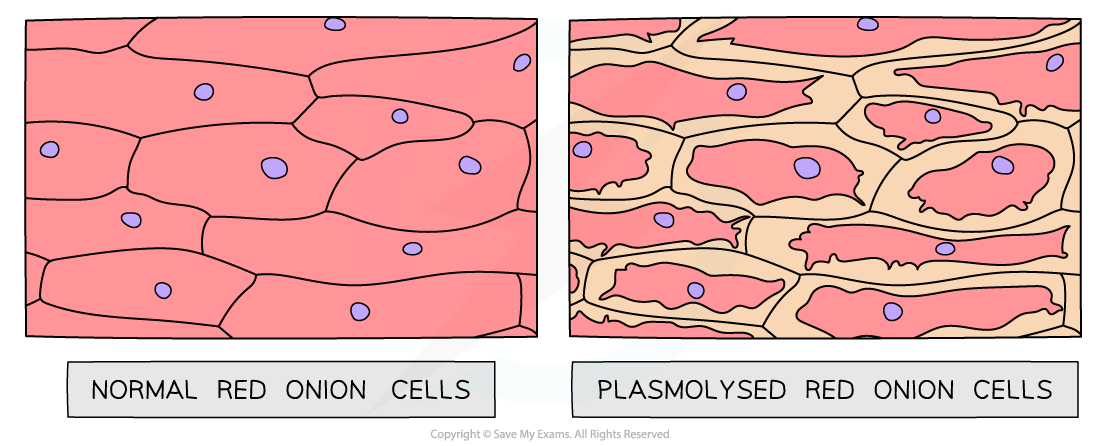
Evidence of osmosis occurring in plant cells can be shown when plant cells undergo plasmolysis:
If a plant cell is placed in a solution with a lower water potential than the plant cell (such as a concentrated sucrose solution), water will leave the plant cell through its selectively permeable cell surface membrane by osmosis
As water leaves the vacuole of the plant cell, the volume of the plant cell decreases
The protoplast (living part of the cell inside the cell wall) gradually shrinks and no longer exerts pressure on the cell wall
As the protoplast continues to shrink, it begins to pull away from the cell wall
This process is known as plasmolysis – the plant cell is plasmolysed
This process can be observed using epidermal strips (sections of the very thin outer layer of tissue in plants)
Plants with coloured sap (such as red onion bulbs, rhubarb petioles and red cabbage) make observations easier
The epidermal strips are placed in a range of concentrations of sucrose solution or sodium chloride solution
The strips are then viewed under a light microscope
Plasmolysis may take several minutes to occur

Unlock more, it's free!
Join the 100,000+ Students that ❤️ Save My Exams
the (exam) results speak for themselves:

Was this revision note helpful?
