1
1 mark
Which one of these causes plantar flexion at the ankle?
Gastrocnemius
Hamstrings
Quadriceps
Tibialis anterior
Was this exam question helpful?
Exam code: 8582
Which one of these causes plantar flexion at the ankle?
Gastrocnemius
Hamstrings
Quadriceps
Tibialis anterior
Was this exam question helpful?
Which bones are found at the shoulder joint?
Femur and tibia
Humerus and radius
Scapula and humerus
Tibia and fibula
Was this exam question helpful?
Which bones are found at the elbow joint?
Femur and tibia
Humerus and radius
Scapula and humerus
Tibia and fibula
Was this exam question helpful?
Figure 1 Shows a young athlete running. The running action involves the use of many joints within the body.
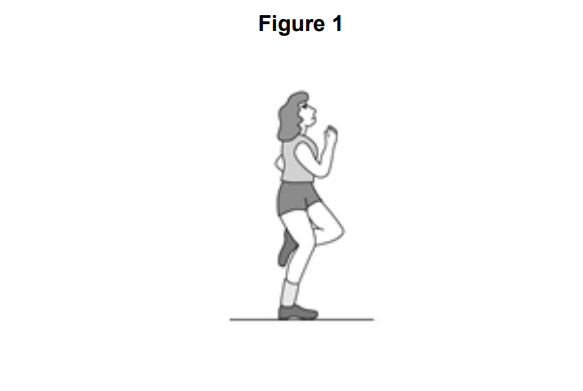
Identify the type of synovial joint working at the shoulder.
Outline how two of the features of the shoulder joint aim to prevent injury occurring
Was this exam question helpful?
Figure 3 shows a person kicking a football.
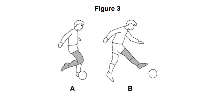
Complete Table 1 to show the joint action occurring at the knee from position A to position B and the agonist muscle group that causes this action.
Table 1
Joint action | Agonist muscle group |
|---|---|
Was this exam question helpful?
Which one of these is the main function of a flat bone?
Allow movement
Blood cell production
Mineral storage
Protection of vital organs
Was this exam question helpful?
Name two major muscles that allow the foot to move at the ankle.
Name two bones found at the elbow.
Name the type of synovial joint at the elbow.
Name three structures of a synovial joint that help to prevent injury
Was this exam question helpful?
Name the type of joint where circumduction can take place.
Was this exam question helpful?
Which one of these structures attaches muscles to bones?
Cartilage
Ligaments
Membranes
Tendons
Was this exam question helpful?
Which one of these muscles is found in the leg?
Deltoid
Gastrocnemius
Latissimus dorsi
Rotator cuffs
Was this exam question helpful?
Which one of these describes an isometric contraction?
The muscle expands in size
The muscle increases in length
The muscle remains the same length
The muscle decreases in length
Was this exam question helpful?
Name the type of joint where abduction can take place.
Was this exam question helpful?
Figure 7 shows a dancer performing a pirouette, balancing on the toes of one foot.
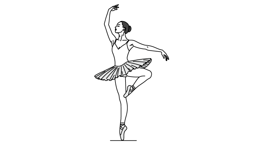
Identify the joint action taking place at the ankle of the supporting foot.
Name the bone found at the front of the knee joint of the supporting leg.
State two functions of the skeleton that are important for the dancer during the pirouette
Was this exam question helpful?
Which one of these joints allows the greatest range of movement?
Hinge joint at the knee
Ball and socket joint at the hip
Hinge joint at the elbow
Pivot joint at the neck
Was this exam question helpful?
Liam is a footballer. During a match he bends his knee to control the ball and then kicks it towards the goal.
Name the type of synovial joint at the knee.
Name two bones found at the knee joint.
Name the bone that sits at the front of the knee joint.
Identify the joint action taking place at the knee as Liam bends his leg to control the ball.
Identify the joint action taking place at the knee as Liam straightens his leg to kick the ball.
Was this exam question helpful?
Flat bones provide a protective function within the body.
Name two flat bones and, using a sporting action of your choice, suggest how these bones provide protection during performance.
Was this exam question helpful?
Before carrying out a weight training session using heavy weights, Robert carries out an appropriate warm up, including stretching of the major muscles that will be used.
Explain what other factors Robert should consider to reduce the chance of injury occurring during the session.
Figure 5 shows a performer weight training. This movement is brought about by the muscular and skeletal systems working together.
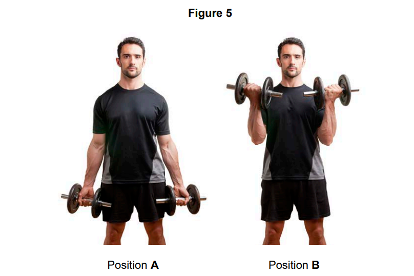
Explain how the muscles and bones work together to produce the movement from position A to position B.
Was this exam question helpful?
Figure 2 shows a person performing a wall sit.
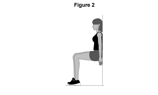
Identify the type of muscular contraction taking place in the legs in Figure 2.
Justify your answer to Question 3(a).
Was this exam question helpful?
Figure 3 shows a basketball player in two different positions (A and B) as they perform the jump shot.
Use Figure 3 to help you answer Questions 13.2 & 13.3
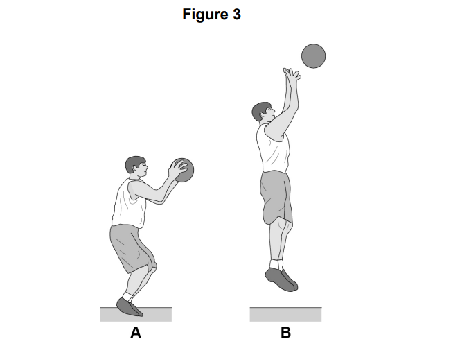
Identify the main agonist at the knee as the basketball player moves from A to B
Identify the type of muscle contraction that is taking place at the knee as the basketball player moves from A to B.
Was this exam question helpful?
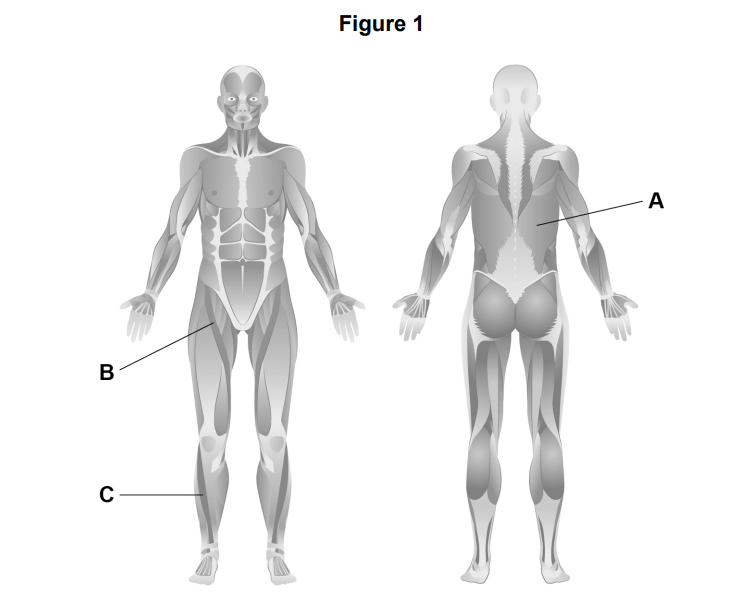
Figure 1 shows a human skeleton.
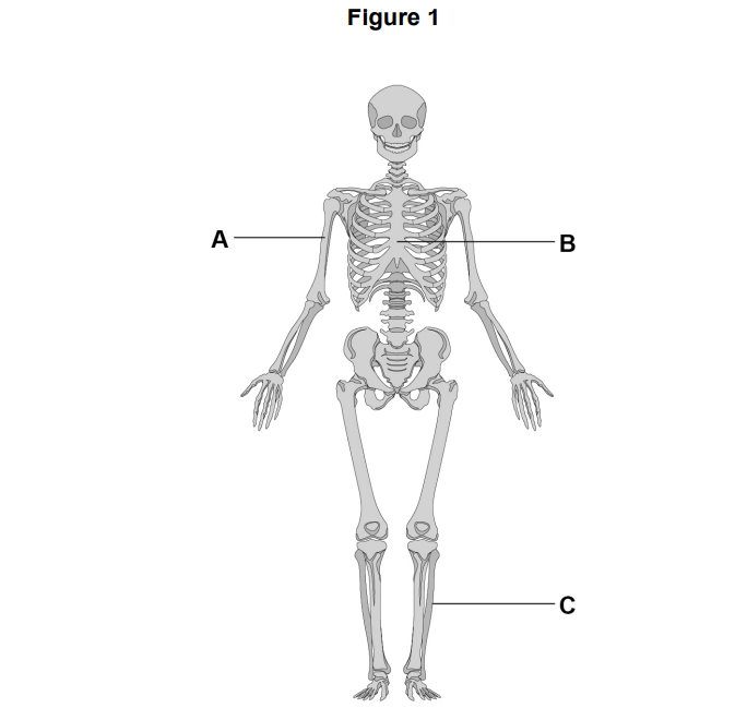
Identify the bones labelled A, B and C in Figure 1.
Was this exam question helpful?
Figure 2 shows an athlete in two different positions (A and B) as the athlete performs a tricep dip.
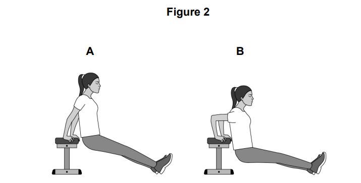
Identify the joint action taking place at the elbow as the arm moves from A to B
Identify the main antagonist at the elbow as the arm moves from A to B.
Identify the type of isotonic muscle contraction that is taking place at the elbow as the arm moves from A to B.
Was this exam question helpful?
Figure 1 shows muscles in the body.
Identify the muscles labelled A, B and C in Figure 1
Name two bones located at the head/neck.
Explain how muscles and bones work to produce movement.
Was this exam question helpful?
Figure 3 shows an individual performing a push-up
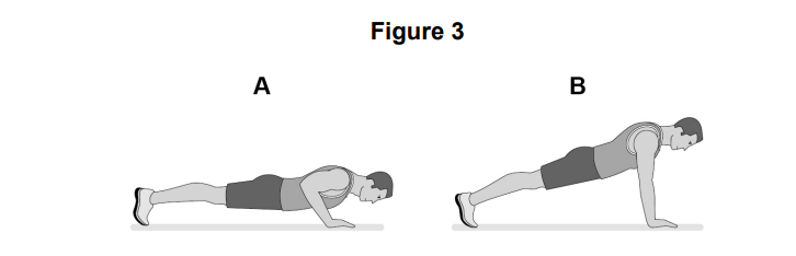
Identify the main agonist at the elbow during the upward phase (A to B) of the push-up.
Identify the type of isotonic muscle contraction taking place at the elbow during the upward phase (A to B) of the push-up.
Was this exam question helpful?
Figure 1 shows a rower at two points in their stroke.
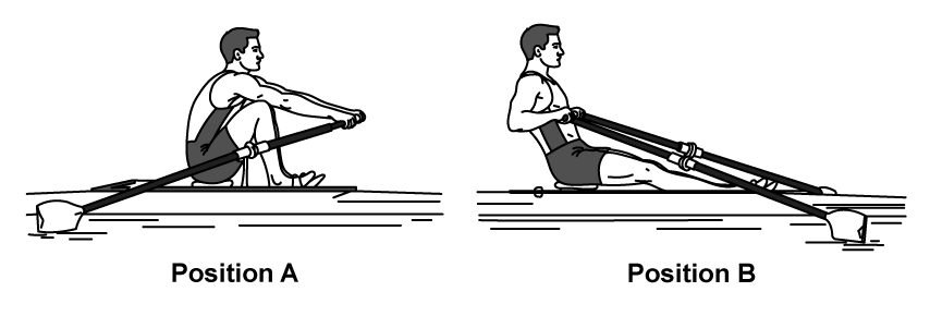
Identify the joint action taking place at the elbow as the rower moves from A to B
Name the main agonist at the elbow during this movement.
The rower then returns to position A by extending their arms back out.
Identify the type of isotonic contraction occurring in the biceps during this return phase and justify your answer
Was this exam question helpful?
Figure 2 shows a gymnast holding a static crucifix position on the rings.
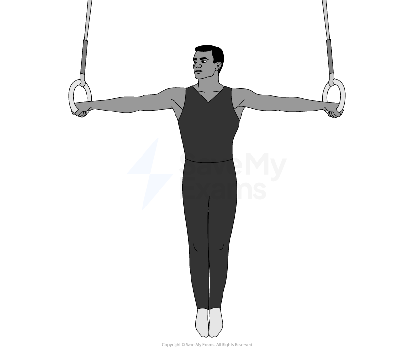
Identify the type of muscular contraction taking place in the shoulders to hold this still position.
Explain why this is an example of the contraction identified in part a.
The gymnast then slowly lowers their arms back down to their sides. Identify the type of isotonic contraction now occurring in the shoulder muscles and justify your answer.
Was this exam question helpful?
A cricket bowler rotates their bowling arm in a full circle to deliver the ball, before following through and decelerating the arm.
Identify the joint action occurring at the shoulder during the full circular bowling action.
Name the type of freely movable joint at the shoulder
Explain why the shoulder joint is capable of this movement but the elbow joint is not.
Explain the role of the rotator cuff muscles for the cricket bowler during the delivery.
Was this exam question helpful?
Figure 4 shows a diagram of a synovial joint with structures labelled A, B, C and D.
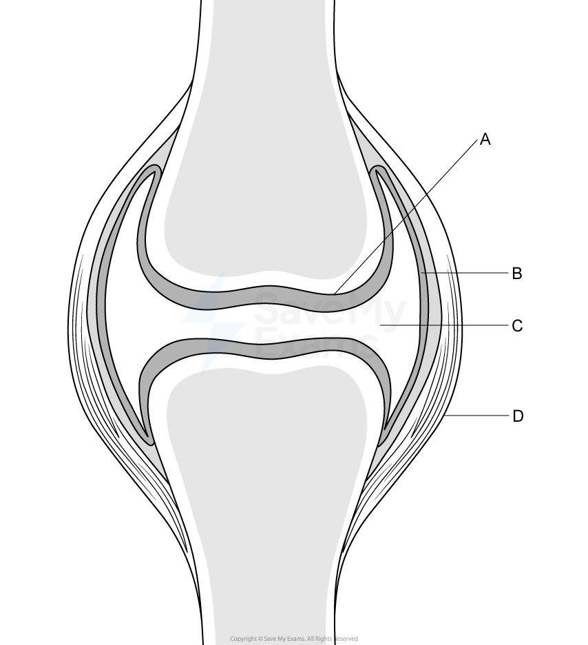
Identify the structures labelled A, B, C and D in Figure 4.
Explain how structure C helps to prevent injury during physical activity.
Name one additional structure found in a synovial joint not shown in Figure 4.
Explain how it helps to prevent injury.
Was this exam question helpful?
Emma is a professional cyclist completing a three-week stage race, cycling for up to six hours per day. By the end of the race her joints are placed under enormous cumulative stress.
Identify two structures of a synovial joint and explain how each structure protects Emma's joints during a six-hour cycling stage.
Emma's coach argues that the skeleton does far more than simply allow movement and that without its other functions Emma could not survive three weeks of racing.
Justify this statement by referring to two functions of the skeleton other than movement
Was this exam question helpful?
Analyse how different types of bones help an individual taking part in a sporting activity of your choice.
Was this exam question helpful?
Tennis players rely heavily on the ball and socket joint at the shoulder to play a variety of powerful shots, such as serves, forehands and backhands.
*Evaluate the importance of the range of movements possible at the shoulder joint for a tennis player during a match
Was this exam question helpful?
Jake is a 16-year-old football player who tears his anterior cruciate ligament (ACL) during a match. His surgeon explains that the ACL connects the femur to the tibia inside the knee joint and prevents the tibia from sliding forward.
Identify the type of connective tissue that has been damaged.
Explain why this injury would prevent Jake from being able to kick a ball effectively.
Jake's physiotherapist says that stronger muscles around the knee joint could have reduced the risk of this injury occurring.
Explain how the muscles acting at the knee help to protect the joint during high-impact movements such as tackling and landing.
Was this exam question helpful?
Figure 6 shows a weightlifter performing a heavy back squat at the bottom of the movement, preparing to push the weight back up.
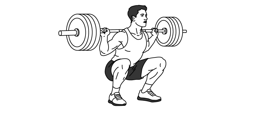
Analyse how the musculoskeletal system enables the weightlifter to perform the upward pushing phase of the squat.
Was this exam question helpful?
Amara is a 400m sprinter. Her coach films her race and analyses her movement at two key moments:
Moment 1: The drive phase out of the blocks, where Amara pushes powerfully off her back leg.
Moment 2: The final 50m, where Amara’s leg muscles suffer from severe fatigue and she struggles to maintain her speed.
Analyse how the skeletal and muscular systems work together at the knee joint to produce the drive phase in Moment 1, and evaluate why her muscles experience severe fatigue during Moment 2.
Was this exam question helpful?