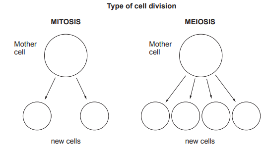
Mitosis and meiosis are the two types of cell division. Image 3 shows the number of new cells formed after mitosis and meiosis in a human cell.
Image 3
Cell division by mitosis can sometimes get out of control. This is what causes cancer. Cancer often begins in one organ of the body before spreading to others.
One in two people will develop some form of cancer. Cancer can occur at any age but it is more likely to occur in the elderly.
Use this information and your own knowledge to answer the following questions.
(i) State how many daughter cells are made during:
[2]
I Mitosis ..........
II Meiosis ..........
(ii) Complete the following table by writing True or False against each statement.
[3]
Statement | True or False |
|---|---|
Both types of cell division can cause cancer. | |
Cancer is more likely to occur the older you are. | |
The number of chromosomes in each cell is the same after mitosis and meiosis. | |
Both types of cell division occur all over the body. |
Was this exam question helpful?
![![[Figure: Image 3.1 — a diagram of an animal cell showing the cell membrane, cytoplasm and nucleus, unlabelled]](https://cdn.savemyexams.com/uploads/2026/08/37359-3-1.png){width=251 height=218}](https://cdn.savemyexams.com/cdn-cgi/image/f=auto,width=3840/https://cdn.savemyexams.com/uploads/2026/08/39911-3-1.png)
![![Figure: karyotype image showing a complete set of paired chromosomes from a cell, arranged in order by size, with the sex chromosome pair positioned at bottom right]](https://cdn.savemyexams.com/cdn-cgi/image/f=auto,width=3840/https://cdn.savemyexams.com/uploads/2026/08/35692-3-1.png)
![[Figure: diagram showing a stem cell undergoing mitosis then differentiating into multiple tissue types: bone, muscle, cartilage, and fat]](https://cdn.savemyexams.com/cdn-cgi/image/f=auto,width=3840/https://cdn.savemyexams.com/uploads/2026/08/13120-3-2.png)
![[Figure: Poster titled 'Human Stem Cell Differentiation' showing stem cells differentiating into intestinal cells, liver cells, cardiac cells, nerve cells, blood cells, and muscle cells]](https://cdn.savemyexams.com/cdn-cgi/image/f=auto,width=3840/https://cdn.savemyexams.com/uploads/2026/08/44908-2a.png)
![[Figure: Diagram showing a body cell from the ovary of a cat (38 chromosomes) undergoing cell division P to produce egg cell A; a sperm cell from a cat (cell B) combines with cell A during fertilisation to produce zygote C; cell C then undergoes cell division Q to produce cell D]](https://cdn.savemyexams.com/cdn-cgi/image/f=auto,width=3840/https://cdn.savemyexams.com/uploads/2026/08/62849-3a.png)
![[Figure: Diagram A shows a starting cell with 4 chromosomes dividing to produce two daughter cells, each with 4 chromosomes (identical to the start). Diagram B shows a starting cell with 4 chromosomes dividing to produce four daughter cells, each with 2 chromosomes]](https://cdn.savemyexams.com/cdn-cgi/image/f=auto,width=3840/https://cdn.savemyexams.com/uploads/2026/08/22561-6a.png)