Cell Division & Stem Cells (WJEC GCSE Science (Double Award): Biology): Exam Questions

Exam code: 3430

41 mins9 questions
1
5 marks

Mitosis and meiosis are the two types of cell division. Image 3 shows the number of new cells formed after mitosis and meiosis in a human cell.

Image 3
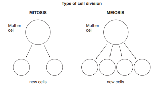
Diagram comparing cell division: mitosis shows one mother cell forming two new cells, while meiosis shows one mother cell forming four new cells.

Cell division by mitosis can sometimes get out of control. This is what causes cancer. Cancer often begins in one organ of the body before spreading to others.

One in two people will develop some form of cancer. Cancer can occur at any age but it is more likely to occur in the elderly.

Use this information and your own knowledge to answer the following questions.

(i) State how many daughter cells are made during:

[2]

I Mitosis ..........

II Meiosis ..........

(ii) Complete the following table by writing True or False against each statement.

[3]

Statement

True or False

Both types of cell division can cause cancer.

Cancer is more likely to occur the older you are.

The number of chromosomes in each cell is the same after mitosis and meiosis.

Both types of cell division occur all over the body.

2
1 mark

Image 3.1 shows an animal cell. Draw an arrow to show the structure where chromosomes are found.

Image 3.1

![[Figure: Image 3.1 — a diagram of an animal cell showing the cell membrane, cytoplasm and nucleus, unlabelled]](https://cdn.savemyexams.com/uploads/2026/08/37359-3-1.png){width=251 height=218}
3
6 marks

During both mitosis and meiosis, a single cell divides into a number of cells.

Give a description of both types of cell division by stating where each type of cell division occurs, the number of cells produced and the functions of both types of cell division.

Diagrams will not be credited.

4a
5 marks

Image 3.1 shows a complete set of chromosomes from the cell of an organism.

Image 3.1

![Figure: karyotype image showing a complete set of paired chromosomes from a cell, arranged in order by size, with the sex chromosome pair positioned at bottom right]

(i) State the evidence, shown in Image 3.1, that suggests that the set of chromosomes may have come from a human cell.

[1]

(ii) Suggest a reason why it is not possible to be certain that the set of chromosomes came from a human cell.

[1]

(iii)

I. On Image 3.1, draw a circle around the sex chromosomes.

[1]

II. State the sex of the organism from which these chromosomes were taken.

[1]

..........

(iv) State how Image 3.1 would differ if the cell from this organism had been produced by meiosis.

[1]

4b
1 mark

State the disease caused by uncontrolled mitosis.

4c
4 marks

Image 3.2 shows some information about human cells.

Image 3.2

[Figure: diagram showing a stem cell undergoing mitosis then differentiating into multiple tissue types: bone, muscle, cartilage, and fat]

(i) Using the information from Image 3.2, explain what is meant by the term stem cell.

[1]

(ii) Researchers at the Washington State University are working on stem cells to transform them into insulin-secreting pancreas cells. They hope that these cells can be used to cure diabetes. However, they have been unable to regulate how much insulin these new pancreas cells produce. Sometimes they produce too much and other times they produce too little.

Describe the effect of the new pancreas cells producing too much insulin in the body.

[3]

5
1 mark

Melanoma is a type of skin cancer. Briefly describe how cancer occurs in the body.

6
1 mark

The surgeons are using stem cells taken from the patient to replace damaged heart muscle.

Suggest one benefit of using stem cells from the patient rather than from a donor.

7a
1 mark

The poster is about some uses of stem cells.

[Figure: Poster titled 'Human Stem Cell Differentiation' showing stem cells differentiating into intestinal cells, liver cells, cardiac cells, nerve cells, blood cells, and muscle cells]

Stem cells can differentiate.

Using one example from the poster, explain the meaning of differentiate.

7b
3 marks

Stem cells undergo rapid mitosis.

The table gives five features of cell division. Some features relate to mitosis and some relate to meiosis.

Complete the table by writing true or false in each row to show the features that relate only to mitosis.

Feature of cell division

True or False

daughter cells are genetically identical

............................................

produces four daughter cells

............................................

daughter cells retain the original chromosome number

............................................

daughter cells have chromosomes in pairs

............................................

produces gametes

............................................

7c
3 marks

A patient receiving stem cell therapy may be treated with cells taken from their own body or from embryos.

Give three advantages of the use of stem cells taken from a patient compared with those from an embryo.

8
4 marks

The diagram shows the two types of cell division occurring in a cat and the process of fertilisation. The number of chromosomes in a body cell from the ovary of a cat is also shown.

[Figure: Diagram showing a body cell from the ovary of a cat (38 chromosomes) undergoing cell division P to produce egg cell A; a sperm cell from a cat (cell B) combines with cell A during fertilisation to produce zygote C; cell C then undergoes cell division Q to produce cell D]

(i) State the type of cell division occurring at points:

P ....................................................................................

Q ....................................................................................

[1]

(ii) State the number of chromosomes in the following cells:

A .........................................

B .........................................

C .........................................

D .........................................

[2]

(iii) Apart from chromosome number, state one other difference, between the daughter cells produced by cell division P and cell division Q.

9
6 marks

Mitosis and meiosis are two types of cell division.

Diagrams A and B show the start and result of both types of cell division in a cell with four chromosomes.

[Figure: Diagram A shows a starting cell with 4 chromosomes dividing to produce two daughter cells, each with 4 chromosomes (identical to the start). Diagram B shows a starting cell with 4 chromosomes dividing to produce four daughter cells, each with 2 chromosomes]

Use the diagrams and your own knowledge to describe the two types of cell division.

You should:

  • state which diagram shows mitosis and which shows meiosis

  • describe what you can see at the start and the result for each type of cell division

  • give one function for each type of cell division