The Visual Sensory System (College Board AP® Psychology): Study Guide

Structures and functions of the visual sensory system
Light enters the eye and is processed in a sequence of structures before reaching the brain:
Light first passes through the cornea
This is a transparent, protective outer layer that begins to focus incoming light
It then passes through the pupil
This is the opening in the centre of the eye that controls how much light enters
The iris (the colored part of the eye) controls the size of the pupil
In bright light the pupil shrinks, in dim light it widens
Light then passes through the lens
The lens fine-tunes the focus by changing shape
Finally, light hits the retina
This is the photosensitive surface at the back of the eye where transduction occurs
Accommodation is the process by which the lens changes shape
It curves more or less to focus light from objects at different distances onto the retina
E.g., your lens flattens when you look at a distant object and curves more sharply when you read a book up close
When accommodation is insufficient, the result is one of the following:
nearsightedness or myopia - difficulty seeing distant objects
farsightedness or hyperopia - difficulty seeing close objects
The fovea is the central region of the retina containing the highest concentration of cones
E.g., when you look directly at a word on this page, you are focusing it onto your fovea
This is why peripheral vision is blurrier than central vision
The blind spot is the point where the optic nerve exits the eye
There are no photoreceptors here, so no visual information is captured
The brain fills in the gap using surrounding visual information, so the blind spot is not consciously perceived under normal conditions
E.g., you do not notice a black hole in your visual field even though one technically exists

Rods and cones
The retina contains two types of photoreceptor cells, rods and cones, which have different functions:
Rods | Cones | |
|---|---|---|
Location | Periphery of the retina. | Concentrated in the fovea. |
Function | Detect shapes, movement, and low-light stimuli. | Detect color and fine detail. |
Color | Cannot detect color. | Three types: blue (short wavelength), green (medium), red (long wavelength). |
Conditions | Active in dim/low-light environments. | Require bright light. |
Adaptation | Responsible for dark adaptation (~20–30 min). | Responsible for light adaptation (fast). |
Example | When you walk into a dark movie theater from bright sunlight, you initially cannot see — your rods are gradually becoming more sensitive. After 20–30 minutes, dark adaptation is complete. | When you walk out of the theater into bright sunlight, your cones take over almost immediately — light adaptation is much faster than dark adaptation. |
Ganglion cells are the output neurons of the retina
Their axons bundle together to form the optic nerve, which carries visual signals to the thalamus and then to the occipital lobe for processing
Theories of color vision
Two theories work together to explain how we see color:
Trichromatic theory
Opponent-process theory
Trichromatic theory
Trichromatic theory (Young-Helmholtz) suggests that the retina contains three types of color receptors
blue
green, and
red cones
All colors are produced by combinations of activation across these three
E.g., mixing red and green cone activation produces the perception of yellow
This theory explains color perception at the receptor/cone level
Opponent-process theory
Opponent-process theory argues that color vision is processed in opposing pairs in ganglion cells:
red/green
blue/yellow, and
black/white
When one color in a pair is activated, its opponent is inhibited
E.g., there is no such thing as "reddish-green" because red and green are opponents — activating one suppresses the other
This theory explains color perception at the ganglion cell level, further along the visual pathway
Both theories are needed to explain vision as:
trichromatic theory explains how cones work
opponent-process theory explains what happens next as signals are processed further
Afterimages occur when prolonged staring at a color fatigues those ganglion cells
When you look at a white surface, the opponent process generates the complementary color
E.g., staring at a red image for 30 seconds then looking at a white wall produces a green afterimage — this supports opponent-process theory
Color vision deficiency
When one or more cone types or their corresponding ganglion cells are damaged or irregular, color vision deficiency results
This means that the person cannot perceive the full range of colors
There are two types of deficiency, differing in severity:
Term | Description | Cause | Example |
|---|---|---|---|
Dichromatism | Cannot distinguish along the red/green or blue/yellow continuum. | Damage or irregularity to one cone type or corresponding ganglion cells; sex-linked, more common in males. | A person with red/green dichromatism may struggle to distinguish a ripe red tomato from unripe green ones against a leafy background. |
Monochromatism | Sees only in shades of gray. | Damage to two or more cone types; very rare. | A person sees the world the way a black-and-white photograph looks. A bright yellow banana and a deep red apple would appear as two different shades of gray. |
Brain processing and visual disorders
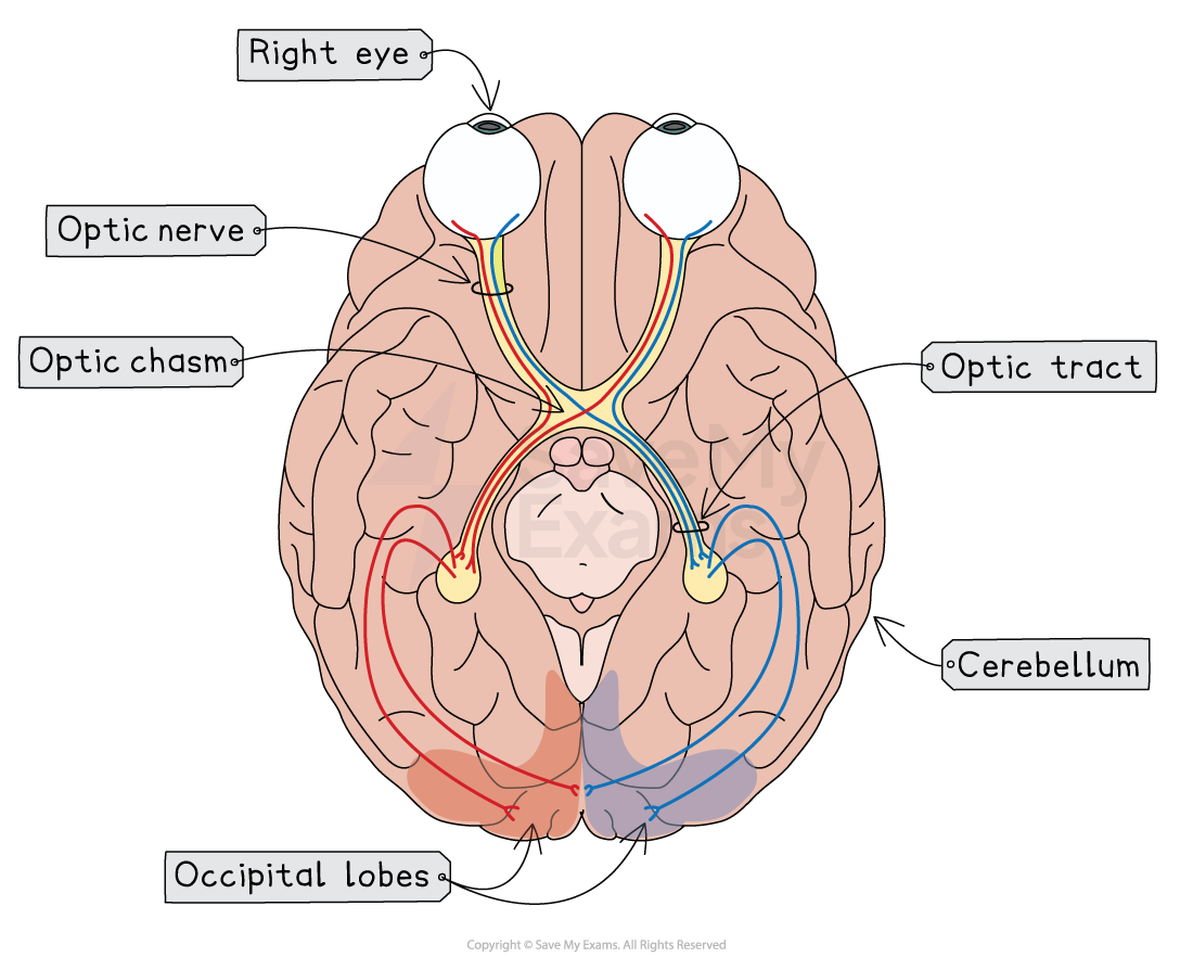
After transduction, signals pass from photoreceptors to bipolar cells, then to ganglion cells, and finally along the optic nerve to the brain
At the optic chiasm, the optic nerves from each eye cross:
Information from the left visual field of both eyes travels to the right hemisphere
Information from the right visual field of both eyes travels to the left hemisphere
Visual processing occurs primarily in the occipital lobe, via two processing streams:
Ventral stream - the "what" pathway, which identifies objects
E.g., recognizing a face or reading a word
Dorsal stream - the "where" pathway, which tracks location and movement
E.g., reaching for a moving object
Prosopagnosia, otherwise known as face blindness, is caused by damage mainly to the occipital lobes
A person with prosopagnosia cannot recognize faces even of close family members, and must rely on other cues such as voice, hair, or gait
Blindsight is another condition caused by occipital lobe damage
This is where an individual cannot consciously see but can still respond to visual stimuli
E.g., a person with blindsight may accurately reach for or dodge an object they report not being able to see, which shows that some visual processing occurs outside of conscious awareness
Examiner Tips and Tricks
For Skill 1.A, visual system questions may describe a scenario and ask you to identify the structure or process involved
Work through the visual pathway in order (cornea → pupil → lens → retina → optic nerve → occipital lobe) to locate where the problem is occurring
E.g., difficulty focusing at different distances - the problem is at the lens (accommodation), not the photoreceptors
E.g., loss of color vision - the problem is at the cones or their corresponding ganglion cells
E.g., inability to recognize faces - the problem is at the occipital/temporal lobe (prosopagnosia)
For Skill 2.B, research on color vision deficiency is typically non-experimental, as researchers cannot randomly assign participants to have or not have color vision deficiency
Be prepared to evaluate why this limits conclusions about cause and effect and why case studies and natural experiments are used instead
The CED explicitly names dichromatism and monochromatism
Be prepared to distinguish between them and link each to the specific cone or ganglion cell damage involved (Skill 1.A)

Unlock more, it's free!
Join the 100,000+ Students that ❤️ Save My Exams
the (exam) results speak for themselves:

Was this revision note helpful?