Neuroanatomy (College Board AP® Psychology): Study Guide

The structure & function of neurons
Neurons are individual nerve cells that transmit information throughout the nervous system
They are the fundamental building blocks of all behavior and mental processes
The human nervous system contains billions of neurons that communicate with one another via electrical and chemical signals
Neurons fire in only one direction — from dendrites through the cell body and down the axon toward the next neuron
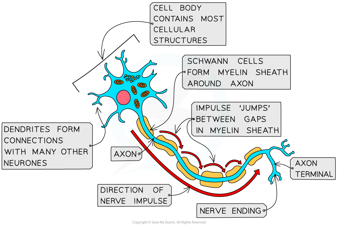
Structure of a typical neuron
Dendrites are branch-like extensions that receive incoming signals from other neurons and transmit them toward the cell body
The cell body (soma) contains the nucleus and sustains the life of the neuron
It also integrates incoming signals from the dendrites
The axon is a long, tube-like structure that carries the electrical signal away from the cell body toward the terminal buttons
The myelin sheath is a fatty coating surrounding the axon of some neurons that insulates the axon and speeds up the transmission of electrical signals
Gaps in the myelin sheath, known as nodes of Ranvier, further accelerate signal transmission
When the myelin sheath deteriorates — as in the disorder multiple sclerosis — signal transmission slows or fails, disrupting movement and sensation
The terminal buttons are branched endings at the tip of the axon that release chemical messengers (neurotransmitters) into the gap between neurons
The synapse is the gap between the terminal buttons of one neuron and the dendrites of the next neuron, across which chemical communication occurs
Glial cells
Glial cells are non-neuronal cells found throughout the nervous system that support the function of neurons
Glial cells provide:
structure — forming the physical scaffolding that holds neurons in place
insulation — producing the myelin sheath that coats axons
communication — facilitating signaling between neurons
waste transport — clearing debris and dead cells from the nervous system
Glial cells do not transmit information themselves but are essential to the functioning of neurons
Without them, neural communication would break down
Glial cells and neurons together form the basis of the nervous system and are the building blocks of all behavior and mental processes
Three types of neurons
Three types of neurons work together in the nervous system, each with a distinct function:
Sensory neurons
Interneurons
Motor neurons
Neuron type | Also called | Direction of signal | Function |
|---|---|---|---|
Sensory neurons | Afferent neurons | PNS → CNS | Carry information from sensory receptors toward the brain and spinal cord |
Interneurons | Relay neurons | Within CNS | Connect sensory and motor neurons; process and relay signals within the CNS |
Motor neurons | Efferent neurons | CNS → PNS | Carry instructions from the brain and spinal cord out to muscles and organs |

The reflex arc
The reflex arc demonstrates how sensory, motor, and interneurons work together within the CNS and PNS to produce a rapid, automatic response to a stimulus
The brain does not need to be involved for the reflex to occur, as the spinal cord coordinates the response and informs the brain afterward
The reflex arc is an adaptive response
Its speed protects the body from harm
The sequence of a reflex arc:
A sensory receptor detects a stimulus, e.g. intense heat from touching a hot surface
A sensory neuron carries the signal from the receptor toward the spinal cord
An interneuron in the spinal cord processes the signal and relays it to the appropriate motor neuron
A motor neuron carries the signal from the spinal cord out to the relevant muscle
The muscle contracts, producing the reflex response, e.g. pulling the hand away
Conscious awareness of the stimulus (e.g. feeling pain) follows the reflex response
The brain is informed after the protective action has already occurred
The reflex arc illustrates how the CNS and PNS work together
the spinal cord (CNS) coordinates the response while sensory and motor neurons (PNS) carry signals to and from the body
Examiner Tips and Tricks
The AP exam tests the function of neurons and glial cells, not their structure (Skill 1.A)
You will not be asked to label parts of a neuron, but you will need to explain what each part does and how it relates to behavior
E.g. how myelin sheath damage in multiple sclerosis disrupts neural communication
If a scenario describes a rapid automatic response to a stimulus, identify it as a reflex arc and explain which neuron type is responsible for each stage (Skill 1.A)
You may be shown a diagram of a neuron or reflex arc and asked to identify a concept (Skill 3.A)
Make sure you can recognize sensory, motor, and interneurons by their direction of signal, not just by name

Unlock more, it's free!
Join the 100,000+ Students that ❤️ Save My Exams
the (exam) results speak for themselves:

Was this revision note helpful?