PET Scans (OCR A Level Physics): Revision Note
Exam code: H556

Positron Emission Tomography (PET) Scanning
Positron Emission Tomography (PET) is defined as:
A type of nuclear medical procedure that images tissues and organs by measuring the metabolic activity of the cells of body tissues
In PET scanning, a beta-plus emitting radioactive tracer is used in order to stimulate positron-electron annihilation to produce gamma photons
These are then detected using a ring of gamma cameras
Principles of PET Scanning
Before the scan
The patient is injected with a beta-plus emitting isotope, usually fluorine-18 (F-18)
During the scan
The part of the body being studied is surrounded by a ring of gamma cameras
The positrons from the F-18 nuclei annihilate with electrons in the patient
The annihilation of a positron and an electron produces two identical gamma photons travelling in opposite directions
The delay time between these two gamma ray photons is used to determine the location of the annihilation due to the F-18 tracer
Photons that do not arrive within a nanosecond of each other are ignored, since they cannot have come from the same point
After the scan
Computer connected to the gamma cameras detect the signal and an image is formed by the computer
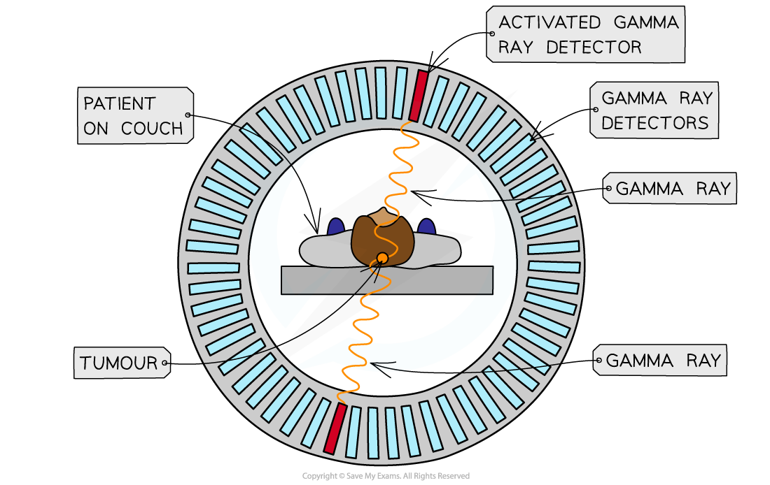
Detecting gamma rays with a PET scanner
Annihilation
When a positron is emitted from a tracer in the body, it travels less than a millimetre before it collides with an electron
The positron and the electron will annihilate, and their mass becomes pure energy in the form of two gamma rays which move apart in opposite directions
Annihilation doesn’t just happen with electrons and positrons, annihilation is defined as:
When a particle meets its equivalent antiparticle they are both destroyed and their mass is converted into energy
As with all collisions, the mass, energy and momentum are conserved
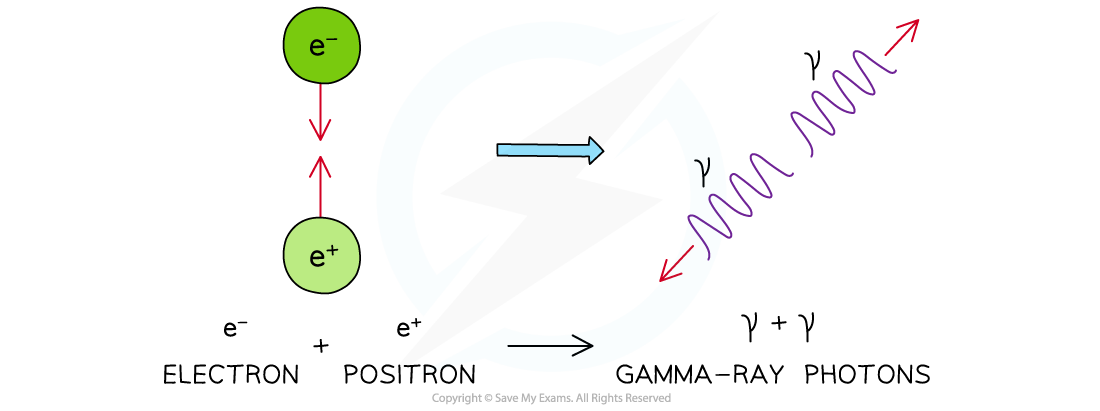
Annihilation of a positron and electron to form two gamma-ray photons
The gamma-ray photons produced have an energy and frequency that is determined solely by the mass-energy of the positron-electron pair
The energy E of the photon is given by
E = hf = mec2
The momentum p of the photon is given by

Where:
me = mass of the electron or positron (kg)
h = Planck's constant (J s)
f = frequency of the photon (Hz)
c = the speed of light in a vacuum (m s–1)
Worked Example
Fluorine-18 decays by β+ emission. The positron emitted collides with an electron and annihilates producing two γ-rays.
(a) Calculate the energy released when a positron and an electron annihilate.
(b) Calculate the frequency of the γ-rays emitted.
(c) Calculate the momentum of one of the γ-rays.
Answer:
Part (a)
Step 1: Write down the known quantities
Mass of an electron = mass of a positron, me = 9.11 × 10⁻³¹ kg
Total mass is equal to the mass of the electron and positron = 2me
Step 2: Write out the equation for mass-energy equivalence
E = mec2
Step 3: Substitute in values and calculate energy E
E = 2 × (9.11 × 10-31) × (3.0 × 108)2 = 1.6 × 10⁻¹³ J
Part (b)
Step 1: Determine the energy of one photon
Planck's constant, h = 6.63 × 10⁻³⁴ J s
Two photons are produced, so, the energy of one photon is equal to half of the total energy from part (a):

Step 2: Write out the equation for the energy of a photon
E = hf
Step 3: Rearrange for frequency f, and calculate

Part (c)
Step 1: Write out the equation for the momentum of a photon

Step 2: Substitute in values and calculate momentum, p

Diagnosis Using PET Scanning
Once the tracer is introduced to the body it has a short half-life, so, it begins emitting positrons (β+) immediately
This allows for a short exposure time to the radiation
A short half-life does mean the patient needs to be scanned quickly and not all hospitals have access to expensive PET scanners
In PET scanning:
Positrons are emitted by the decay of the tracer
They travel a small distance and annihilate when they interact with electrons in the tissue
This annihilation produces a pair of gamma-ray photons which travel in opposite directions
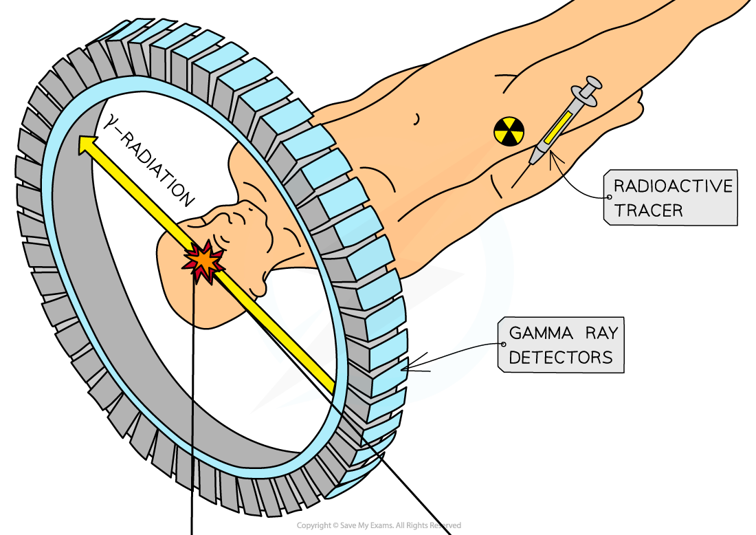
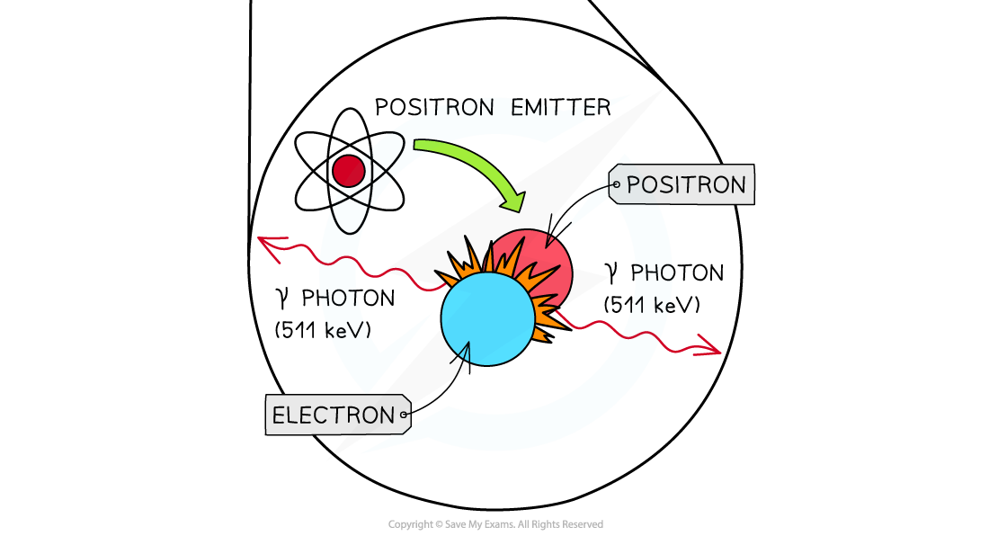
Annihilation of a positron and an electron is the basis of PET Scanning
Image Formation on a Computer
The signals produced by the photomultiplier tubes are used to produce an image
The γ rays travel in straight lines in opposite directions when formed from a positron-electron annihilation
This happens in order to conserve momentum
They hit the detectors in a line – known as the line of response
The tracers will emit lots of γ rays simultaneously, and the computers will use this information to create an image
The more photons from a particular point, the more tracer that is present in the tissue being studied, and this will appear as a bright point on the image
An image of the tracer concentration in the tissue can be created by processing the arrival times of the gamma-ray photons

Unlock more, it's free!
Join the 100,000+ Students that ❤️ Save My Exams
the (exam) results speak for themselves:

Was this revision note helpful?
Build on this topic