The Human Circulatory System (AQA GCSE Combined Science: Synergy: Life & Environmental Sciences): Revision Note
Exam code: 8465

The Heart
The double circulatory system
The human heart is part of a double circulatory system
The circulatory system consists of
Blood vessels
A pump (the heart)
Valves to maintain one-way blood flow
The heart has four chambers divided into two sides
The right side pumps blood to the lungs for gas exchange
The left side pumps blood at high pressure to the body
Benefits of a double circulatory system
Blood loses pressure as it passes through lung capillaries, slowing flow and allowing time for gas exchange
Oxygenated blood returns to the heart so pressure can be increased before being pumped to the body
This allows oxygen to be delivered to body cells more quickly
Heart structure
The right side of the heart receives deoxygenated blood from the body and pumps it to the lungs
Gas exchange occurs in the lungs where oxygen diffuses in and carbon dioxide diffuses out
The left side of the heart receives oxygenated blood from the lungs and pumps it to the body
Veins carry blood towards the heart and arteries carry blood away from the heart
The four chambers are arranged vertically
Atria are the upper chambers
Ventricles are the lower chambers


Examiner Tips and Tricks
The heart is labelled as if it was in the chest so what is left on a diagram is the right-hand side (and vice versa).
You need to know the following structures:
Aorta
Vena cava
Pulmonary artery - the only artery in the body to carry deoxygenated blood
Pulmonary vein - the only vein to carry oxygenated blood
Coronary arteries
Remember arteries carry blood away from the heart, veins towards it.
Knowledge of the names of the heart valves is not required.
Pathway of blood through the heart
Deoxygenated blood enters the right atrium via the vena cava
Blood flows through valves into the right ventricle
The right ventricle pumps blood to the lungs via the pulmonary artery
Oxygenated blood returns to the left atrium via the pulmonary vein
Blood flows through valves into the left ventricle
The left ventricle pumps blood to the body via the aorta
Adaptations of the heart
Ventricles have thicker walls than atria to generate higher pressure
The left ventricle has the thickest wall to pump blood around the body
The right ventricle pumps blood at lower pressure to the lungs
Valves prevent the backflow of blood
The septum separates the two sides of the heart and prevents mixing of oxygenated and deoxygenated blood
Coronary arteries supply the heart muscle with oxygen and glucose for aerobic respiration
Heart rate
Resting heart rate
The natural resting heart rate is controlled by a group of cells located in the right atrium
These cells form a structure called the pacemaker
The role of the pacemaker is to coordinate the contraction of the heart muscle, therefore it regulates the heart rate
Up to a point, the faster the heart contracts, the more quickly oxygenated blood can be delivered around the body
When a person is at rest, the oxygen demand of their cells is relatively low and so a lower heart rate is maintained
When a person is exercising, the oxygen demand of their muscle cells increases so a higher heart rate is necessary
The pacemaker sends out an electrical impulse which spreads to the surrounding muscle cells of the heart, causing them to contract
The pacemaker does this every time the heart needs to “beat”, so if a person has a resting heart rate of 60 beats per minute (bpm), then the pacemaker will be sending out electrical impulses on average once every second
Artificial pacemakers
Sometimes, the pacemaker of the heart stops functioning properly (this can cause an irregular heartbeat)
Artificial pacemakers are electrical devices used to correct irregularities in the heart rate
The device is implanted just under the skin, with a wire that delivers an electrical current to the heart to help it contract regularly
Examiner Tips and Tricks
The pacemaker is located in the wall of the right atrium – you may be asked to locate it on a diagram in the exam
Blood vessels
Types of blood vessels
The body contains three different types of blood vessels:
Arteries: transport blood away from the heart (usually at high pressure)
Veins: transport blood to the heart (usually at low pressure)
Capillaries: links arteries to veins within the tissues of the body
Blood vessels structure
The walls of each type of blood vessel have a structure that relates to the function of the vessel
Blood flows through the lumen of a blood vessel; the size of the lumen varies depending on the type of blood vessel (with arteries having a narrow lumen, and the veins a wider one)
The lumen of the capillaries is extremely narrow, at the smallest the width of a red blood cell!
The structure of arteries, capillaries and veins diagram
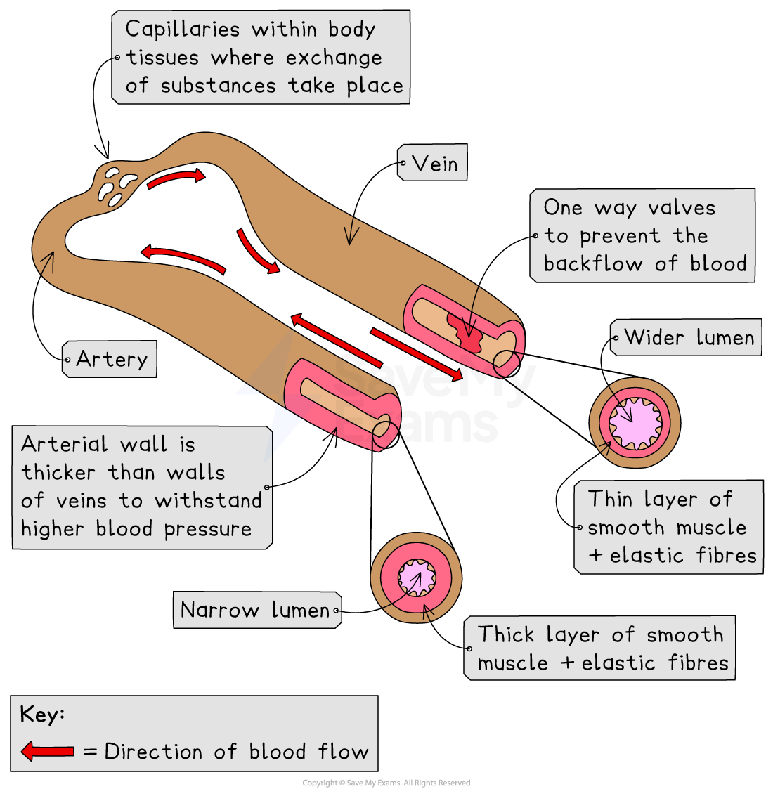
How structure relates to function
Arteries must withstand and maintain high pressures from the contracting and relaxing heart
Their thick walls contain collagen, smooth muscle, and elastic fibers
The elastic fibers allow expansion and recoil, maintaining high blood pressure alongside a narrow lumen
Veins receive low-pressure blood from capillaries and return it to the heart
They have thinner walls with fewer layers of collagen, smooth muscle, and elastic fibers, but a much larger lumen
Veins contain valves to prevent backflow
Capillary walls consist of a single layer of endothelial cells, minimising the diffusion distance for oxygen and carbon dioxide
These walls have pores that allow blood plasma to leak out and form tissue fluid
Examiner Tips and Tricks
Do not confuse the wall of the capillary being ‘one cell thick’ to mean that the cells that form the capillary wall have “cell walls”. Animal cells never have cell walls.
The Lungs & the Circulatory System
The human circulatory system transports substances around the body
It works closely with the gaseous exchange system in the lungs
Blood transports
Oxygen from the lungs to body cells
Carbon dioxide from body cells to the lungs
Dissolved food molecules (such as glucose) from the digestive system to cells
These substances are needed for respiration or removed as waste
Structure of the lungs
Gas exchange takes place in the lungs
Air enters the lungs through the trachea
The trachea splits into two bronchi, one leading to each lung
Bronchi branch into bronchioles which end in alveoli
Alveoli are surrounded by a dense capillary network

Alveoli and gas exchange
Alveoli are specialised for efficient gas exchange
They have a large surface area
Their walls are very thin, giving a short diffusion distance
The surrounding capillary network maintains a steep concentration gradient
Oxygen diffuses from the air in the alveoli into the blood
Carbon dioxide diffuses from the blood into the alveoli
Examiner Tips and Tricks
Make sure that you can identify the trachea, bronchi, alveoli, and capillary network in the lungs, and that you can explain how the lungs are well adapted for gas exchange.

Unlock more, it's free!
Join the 100,000+ Students that ❤️ Save My Exams
the (exam) results speak for themselves:

Was this revision note helpful?
