Cell Specialisation (AQA GCSE Biology): Revision Note
Exam code: 8461

Specialised cells
A specialised cell is a cell that has a structure that aids its specific function within a tissue, an organ or organ system, or the whole organism
This could relate to cell shape or the combination of cellular structures present within the cell
Cells specialise by undergoing a process known as differentiation
Examiner Tips and Tricks
You may be given some information (including an image) about an unfamiliar cell in an exam and asked to explain how it’s able to carry out its function; make sure that your answers always clearly link structure with function.
Factors to consider when looking for cell adaptations:
Does the cell have a shape which increases its surface area or allows it to interact with other cells?
Are there lots of a particular organelle? What is that organelle's job?
Specialised cells in animals
Nerve cells
Function:
Conduction of electrical impulses
Special features that aid function:
Nerve cells are long, so can conduct nerve impulses between different areas of the body
Extensions of the cytoplasm known as dendrites allowing nerve cells to communicate with other cells
The axon is covered with a fatty sheath which speeds up nerve impulse transmission

Muscle cells
Function:
Contraction for movement
Structures that aid function:
Muscle cells have many mitochondria to release energy for contraction
All muscle cells contain protein filaments that can slide over each other to allow muscle contraction

Sperm cells
Function:
Transfer of genetic material to an egg cell for fertilisation
Structures that aid function:
The mid-piece is packed with mitochondria to release energy (via respiration) for the tail
The tail rotates, propelling the sperm cell forward and allowing it to move
The head contains a nucleus with half the normal number of chromosomes, allowing the sperm cell to fuse with an egg cell to restore the normal chromosome number
The acrosome in the head contains digestive enzymes that can break down the outer layer of an egg cell so that the haploid nucleus can enter to fuse with the egg’s nucleus

Specialised cells in plants
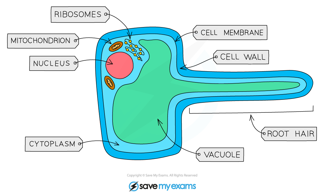
Root hair cells
Function:
Absorption of water and mineral ions from soil
Structures that aid function:
Root hairs increase surface area (SA) so the rate of water uptake by osmosis is greater
Thinner walls than other plant cells so that water can move through easily due to a shorter diffusion distance
Mitochondria release energy for active transport of mineral ions
No chloroplasts. Root hair cells are underground, so they get no light and cannot photosynthesise.
Xylem vessels
Function:
Transport of water and dissolved ions
Structures that aid function:
Xylem is made of dead cells
No end walls between cells to form continuous hollow tubes through which water is passively drawn upwards towards the leaves
Cells contain no organelles or cytoplasm, allowing free passage of water
Outer walls are thickened with a substance called lignin, strengthening the tubes and providing support for the plant

Phloem cells
Function:
Transport of dissolved sugars (and amino acids) from the leaves to the rest of the plant, both up and down the plant (translocation)
Structures that aid function:
Phloem is made of living cells
Cells are joined end-to-end and contain pores in the end walls (sieve plates); this forms tubes which allow sugars and amino acids to flow easily
Cells have very few sub-cellular structures to aid the flow of materials


Unlock more, it's free!
Join the 100,000+ Students that ❤️ Save My Exams
the (exam) results speak for themselves:

Was this revision note helpful?
Build on this topic