Blood Vessels & Blood (AQA GCSE Biology): Revision Note
Exam code: 8461

Blood vessels
The body contains three different types of blood vessels:
arteries: transport blood away from the heart
veins: transport blood to the heart
capillaries: links arteries to veins and are the site of substance exchange with the body cells
Examiner Tips and Tricks
Remember that:
arteries carry blood away from the heart
veins carry blood into the heart
Blood vessel structures
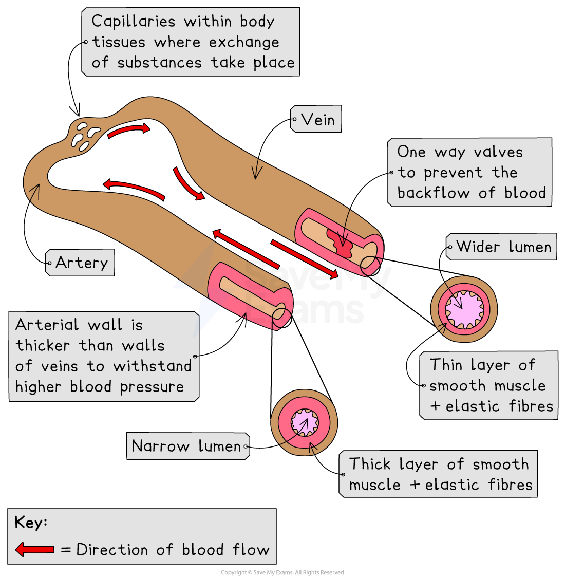
The structure of each type of blood vessel relates to its function
Arteries must withstand and maintain high pressures generated by the contracting heart muscle; features that enable this include:
thick walls that contain muscle for strength and elastic tissue for stretch and recoil
a narrow lumen to maintain high blood pressure
Veins carry a large volume of blood at low-pressure; features that enable this include:
a large lumen to accomodate the large volume of blood
thinner walls, as they do not need to withstand high pressure
valves to prevent backflow of blood
Capillaries carry blood to the cells of the body, supplying required substances and removing waste; features that enable this include:
walls one cell thick, minimising the diffusion distance for exchange
very narrow lumen to slow blood flow, allowing time for exchange to occur
Examiner Tips and Tricks
Never say that capillaries have 'thin cell walls' as this is incorrect; capillaries have walls that are one cell thick.
Rate calculations: Blood flow
The rate of blood flow can be calculated if the volume of blood flow and the time is known
Worked Example
If 2460 ml of blood flows through a blood vessel in 4 minutes, what is the rate of blood flow?
Answer:
Step 1: Rate = volume of blood / number of minutes
= 2460 / 4
= 615 ml/minute
Step 2: From this it may be determined how much blood flows through the same vessel in one hour
= rate of blood flow (ml/min) x 60
= 615 x 60 = 36 900 ml
Structure & function: Blood
The role of blood is to transport useful substances to every cell of the body, and to remove harmful waste substances
It also plays a role in temperature regulation
Blood is a tissue that contains:
plasma
red blood cells
white blood cells
platelets
Components of the blood
Plasma
Plasma is the straw-coloured liquid part of the blood in which the cells are suspended
It transports dissolved substances, including carbon dioxide (from respiring cells to the lungs), glucose and amino acids (from the small intestine to cells), urea (from the liver to the kidneys), hormones, antibodies and ions
It also carries the blood cells and transfers heat around the body
Always name the substance: 'nutrients' and 'waste' do not get the mark
Red blood cells
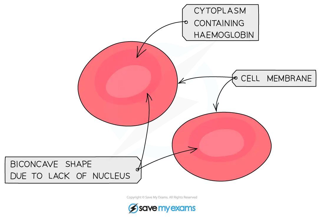
Red blood cells carry oxygen around the body
They have several features that aid their function:
a biconcave disc shape
The biconcave shape gives red blood cells a large surface to volume ratio; this is a key adaptation to maximise the efficiency of diffusion of gases into and out of the cell
haemoglobin
This is a protein that binds to oxygen, allowing it to be transported around the body
no nucleus
This maximises the space available for haemoglobin
White blood cells
White blood cells are part of the immune system, responsible for defending the body from infection by recognising and destroying pathogens
white blood cells defend the body by:
producing antitoxins, which neutralise toxins released by pathogens
Platelets
Platelets are fragments of cells (they contain cytoplasm but no nucleus)
When damage to a blood vessel occurs, the platelets are involved in forming a blood clot to prevent blood loss

Recognising blood cells
An exam question may include a photograph or diagram and ask the identity of the types of blood cell present
Observing and drawing cells under the microscope is an important skill to develop
To identify the cells: white blood cells are larger than red blood cells and are the only blood cells with a nucleus (often lobed); red blood cells are biconcave discs with no nucleus; platelets are much smaller cell fragments with no nucleus


Unlock more, it's free!
Join the 100,000+ Students that ❤️ Save My Exams
the (exam) results speak for themselves:

Was this revision note helpful?
Build on this topic