Required Practical: Microscopy (AQA GCSE Biology): Revision Note
Exam code: 8461

Using a light microscope
Aim: to use a light microscope to observe, draw and label a selection of plant and animal cells, including a magnification scale
Preparing a microscope slide
Specimens must be prepared on a microscope slide to be observed under a light microscope
This must be done carefully to avoid damaging biological specimens
The most common specimens to observe under a light microscope are:
cheek cells (animal cells)
onion cells (plant cells)
Stains are used to highlight structures within cells
Methylene blue can be used to stain cheek cells
Iodine can be used to stain onion cells
To prepare a microscope slide for viewing onion cells:
Remove a thin piece of onion tissue; this can be done by slicing a piece of onion and then using tweezers to peel away the membrane-like tissue on the outside of an onion layer. The layer must be thin so light can pass through it and individual cells can be seen
Place the piece of onion tissue onto a glass slide, ensuring that it is flat and contains no folded areas
Add a drop of iodine stain, which stains the parts of the cell (e.g. nucleus, cytoplasm, cell wall) so they can be seen. Lower a cover slip slowly at an angle to stop air bubbles being trapped; use a paper towel to remove excess stain

Risk assessment:
Iodine solution is an irritant, so wear gloves or wash skin immediately after contact
A sharp knife or scalpel may cut you, so cut on a chopping board, away from your body, keeping fingers away from the blade
Goggles do not protect against cuts, and calling a first aider is not a way to reduce the risk
Viewing a slide under a light microscope
To use a microscope to view a specimen:
clip the slide carefully onto the stage
ensure the lowest-powered objective lens is over the slide
use the coarse adjustment knob to bring the stage up just below the lens
look down the eyepiece and gradually move the stage downwards using the coarse adjustment knob; stop when the image is roughly in focus
bring the image into focus by adjusting the fine-adjustment knob until a clear image is obtained (the focusing knobs make the image sharper; they do not change the magnification, so don't describe them as "zoom")
observe the image with a higher magnification, by changing the objective lens to a higher power and readjust the stage using the coarse and fine adjustment knobs

Biological drawings
Producing biological drawings of specimens viewed under a microscope is a key skill
Biological drawings are scientific, so a set of rules must be followed:
Lines must be drawn with a sharp pencil
Pencil lines should be clear and continuous, with no sketching
No shading should be used
Structures should be drawn as they appear in the field of view
Structures should be in proportion to each other
Features should be clearly labelled
Labels lines should be drawn with a ruler, should clearly touch the item being labelled, and should not cross each other
A scale bar or an indication of the magnification used should be included
Rules for biological drawing
Always draw what you see with a sharp pencil using clear, unbroken lines
All structures drawn should be in proportion
Label all features using straight, uncrossed lines
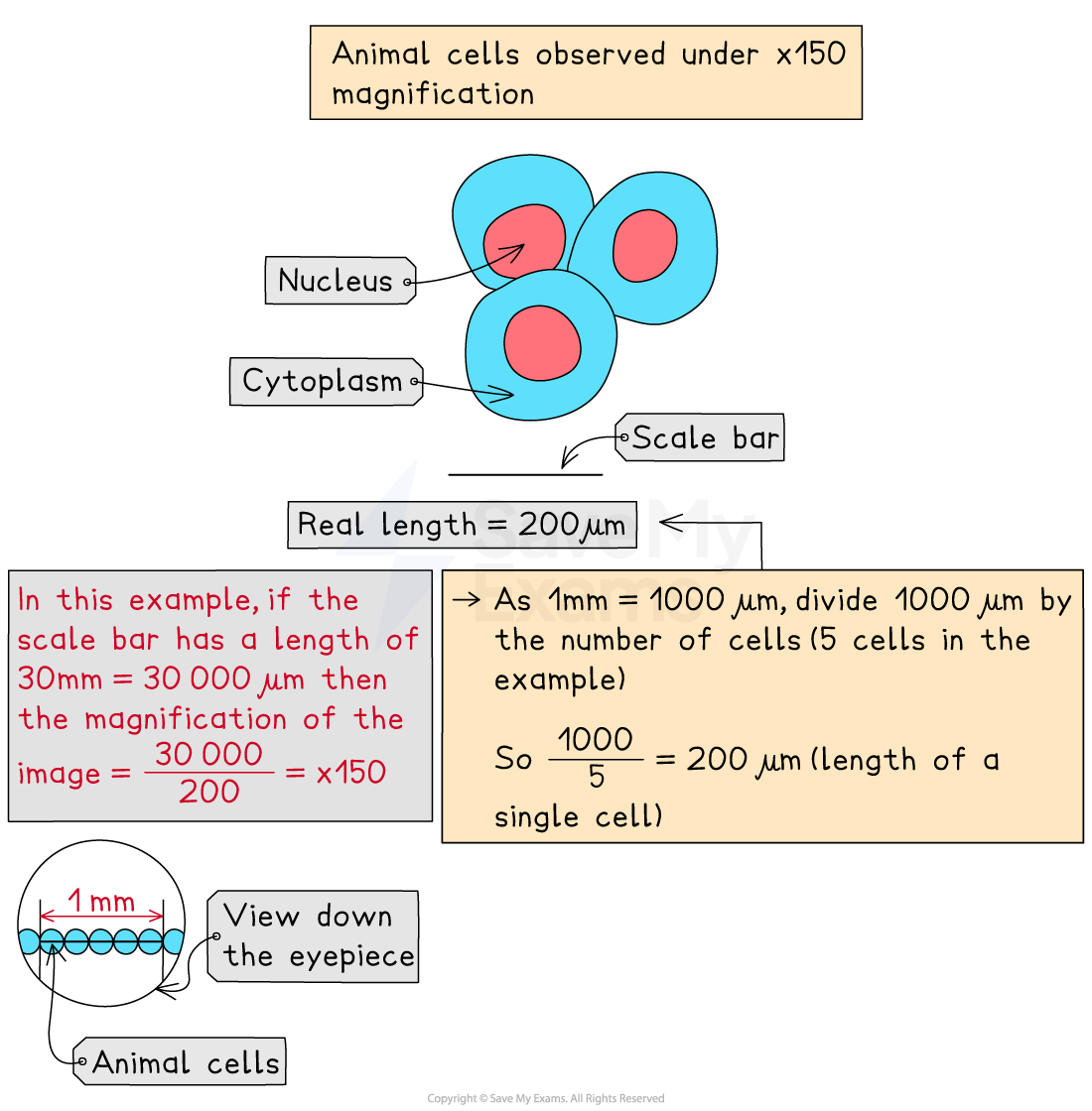
To calculate the size of a single cell
Place a clear plastic ruler on the stage next to the slide
View the ruler and slide under the ×4 or ×10 objective lens and adjust focus to obtain a clear image
Line the cells along 1 mm and count the number of cells that fit across that length
As 1 mm = 1000 µm, divide 1000 µm by the number of cells (5 cells in the example) So 1000 ÷ 5 = 200 µm (length of a single cell)
Estimating the size of cellular features
When cells are viewed under a microscope, it is possible to estimate the relative size or area of the structures inside cells
E.g.when viewing a microscope image:
use a rule to measure the length of two different cell features
calculate how many times larger one feature is than the other

Unlock more, it's free!
Join the 100,000+ Students that ❤️ Save My Exams
the (exam) results speak for themselves:

Was this revision note helpful?
Build on this topic